

Lymphoma is a malignancy arising from lymphocytes or lymphoblasts. It can be restricted to the lymphatic system or arise as extranodal disease. This, along with variable aggressiveness, results in a diverse imaging appearance.
On this page:
Epidemiology
Lymphoma accounts for ~3-4% of all cancers 1-3. They are more common in developed countries.
In children, lymphoma accounts for 10-15% of all cancers, being the third most common form of malignancy 4.
Clinical presentation
Lymphoma can present as nodal or extranodal disease. Hodgkin lymphoma and low-grade non-Hodgkin lymphoma (NHL) classically present as nodal disease, whereas high-grade NHL can present with complications from the mass effects such as superior vena cava obstruction, cauda equina syndrome, etc. Extranodal disease can affect any organ.
Lymphoma often presents with B symptoms (fever, night sweats and weight loss).
Pathology
Lymphomas are malignancies that arise from mature lymphocytes. The aetiology is unknown but potential lymphomatogenic risk factors include 3:
bacterial infection, e.g. Helicobacter pylori
chronic immunosuppression, e.g. post-transplantation
prior chemotherapy (especially alkalising agents) and drug therapy, e.g. digoxin
Classification
Lymphomas are classified according to the WHO classification of tumours of haematopoietic and lymphoid tissues based on cell of origin (e.g. B-cell, T-cell and NK-cell) and then further into numerous other categories and specific diagnoses. The majority (85%) of lymphomas are B-cell with the remainder (15%) being T-cell 3.
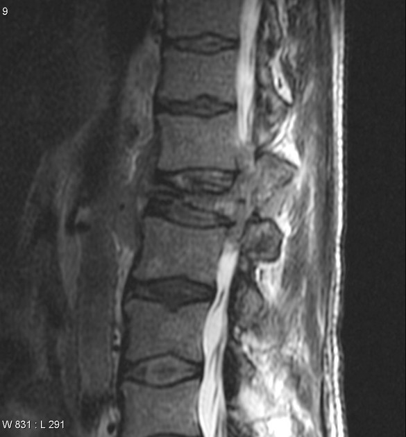
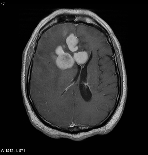
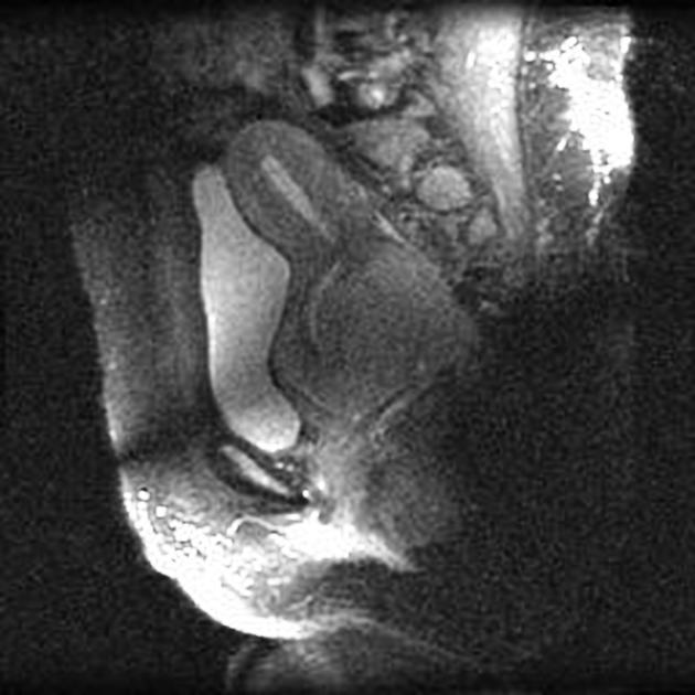
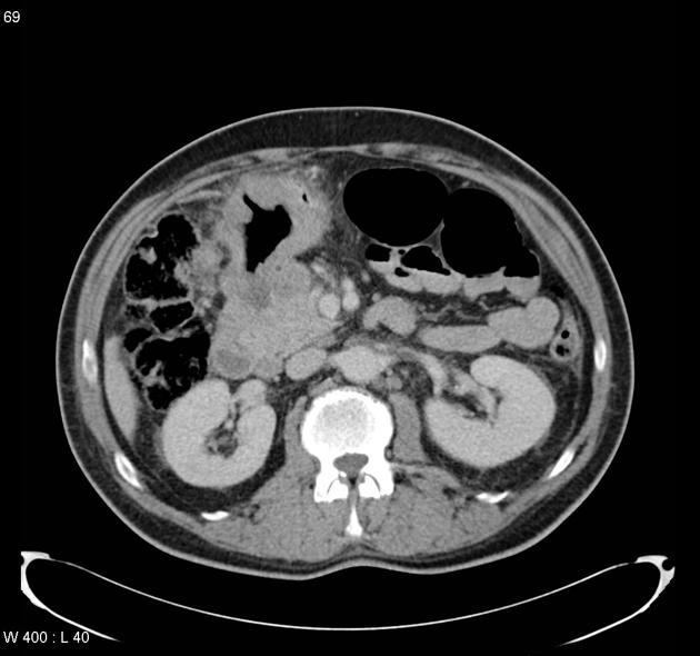
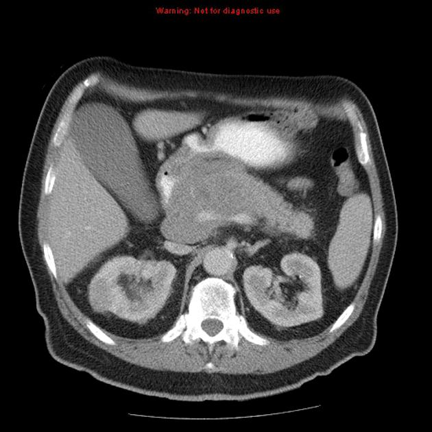
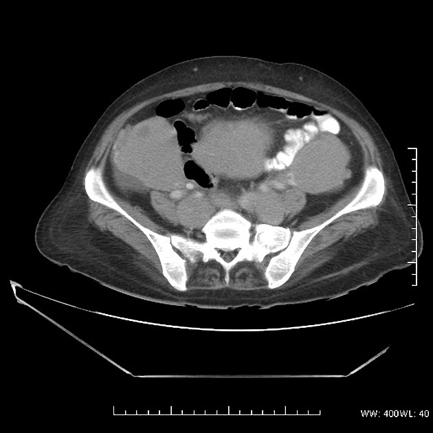
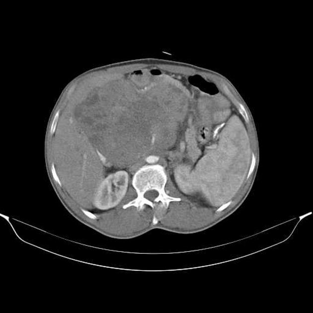
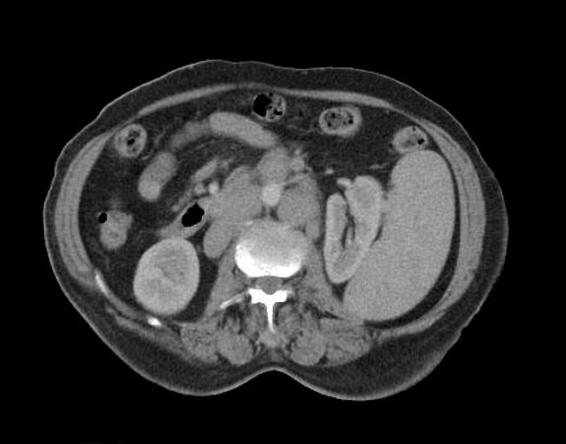
Location
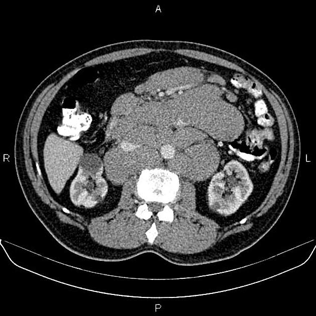
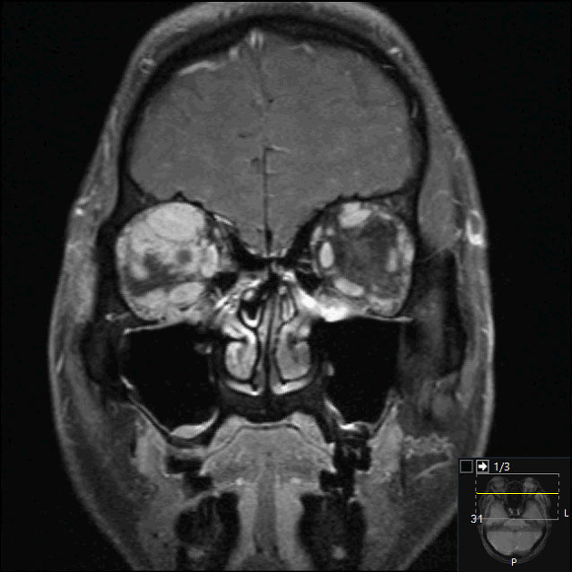
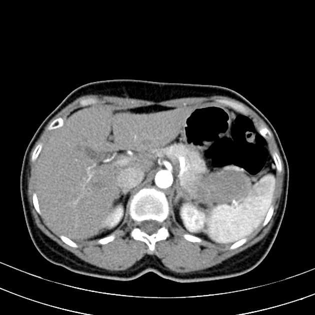
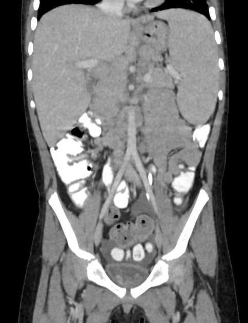
Additionally, it is worth, especially for radiologists, dividing extranodal lymphomas according to the location:
-
central nervous system (CNS)
-
head and neck lymphoma
-
thoracic lymphoma
-
gastrointestinal lymphoma
-
hepatobiliary lymphoma
-
musculoskeletal lymphoma
-
cutaneous lymphoma
-
genitourinary lymphoma
-
multi-regional
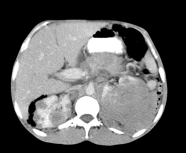
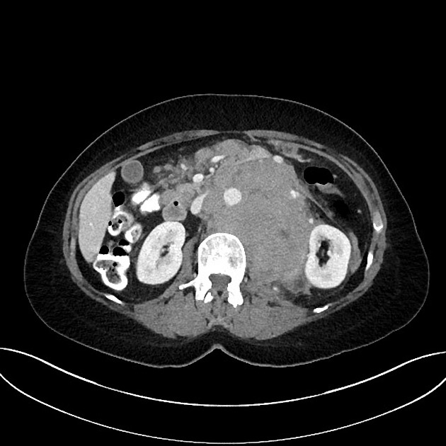
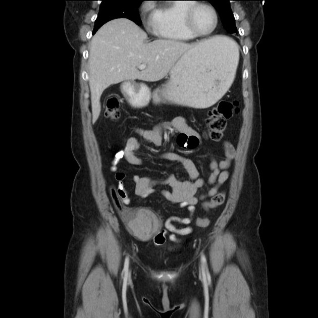
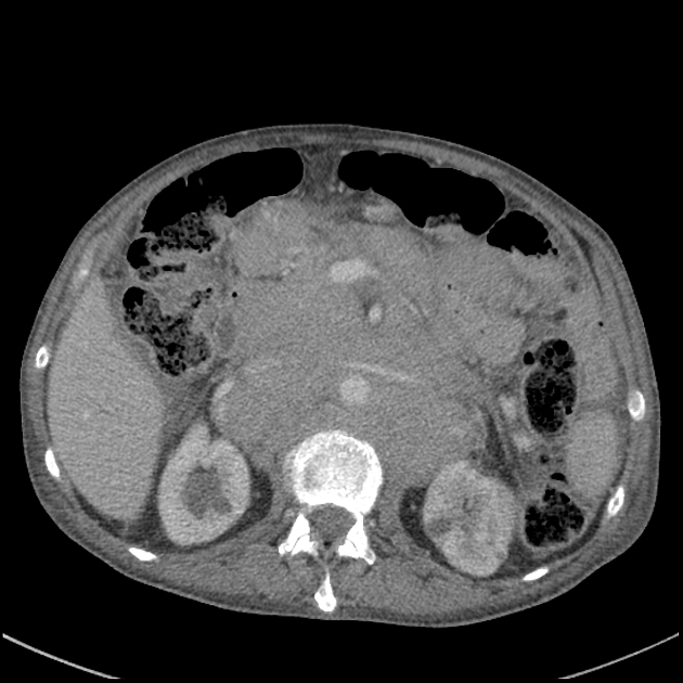
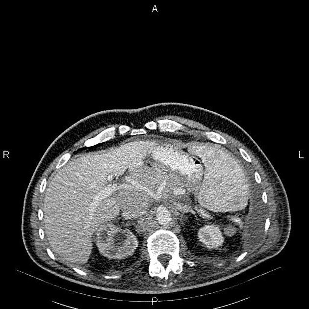
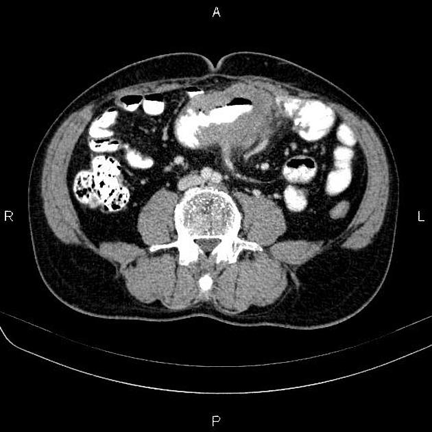
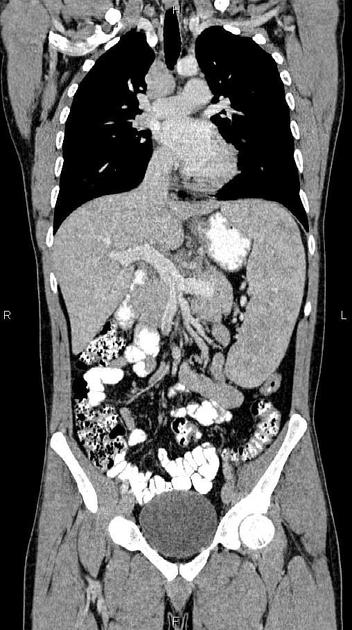
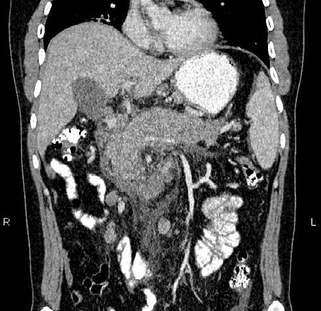
Radiographic features
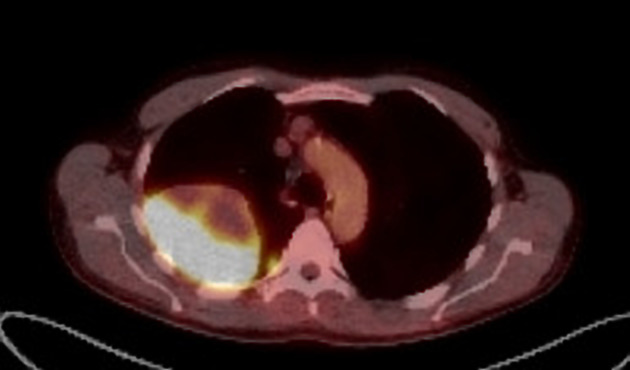
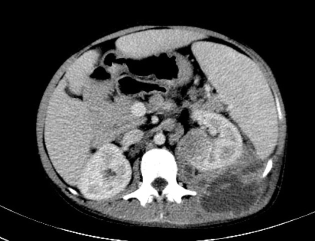
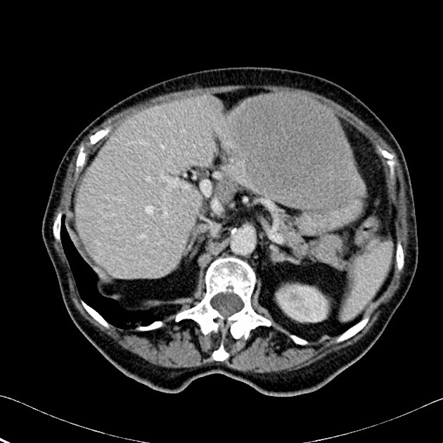
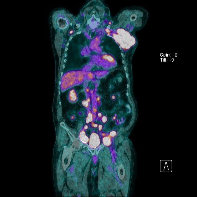
Imaging characteristics will depend on the location and subtype of lymphoma. CT is the workhorse of imaging in lymphoma and plays a crucial role in staging (see main article: lymphoma staging). US and MRI are also used. For example, when assessing cervical lymph nodes (US) or CNS lymphoma (MRI). FDG-PET is used for staging and re-staging of lymphoma.
Treatment and prognosis
Lymphoma cure rates are comparatively high (up to 90%) compared to many other malignancies. Prognosis depends not only on histological subtype and grade but also on stage, hence why imaging plays a pivotal role in treatment. Aggressive lymphomas (e.g. Burkitt lymphoma) typically have a prognosis of weeks without treatment.


 Unable to process the form. Check for errors and try again.