
Ovarian mucinous tumors are a subgroup of ovarian epithelial tumors. They represent 10-15% of all ovarian tumors and ~10% of all malignant ovarian tumors. They are subdivided according to their malignant potential and clinical behavior into:
On this page:
Clinical presentation
Benign mucinous tumors tend to affect women 20-40 years old, whereas borderline and malignant tumors tend to occur in a slightly older age range (40-50 years of age) 6,
Pathology
On histology they show multiple cysts lined by mucinous epithelium, often resembling gastrointestinal-type epithelium 6. KRAS mutations are a common feature.
Subtypes
Mucinous ovarian tumors can be broadly subclassified into three main subgroups:
- ovarian mucinous cystadenoma: ~80 %
- ovarian borderline mucinous tumor: ~10-15 %
- ovarian mucinous cystadenocarcinoma: ~5-10 %
Radiographic features
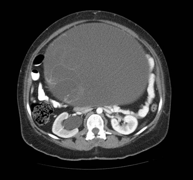
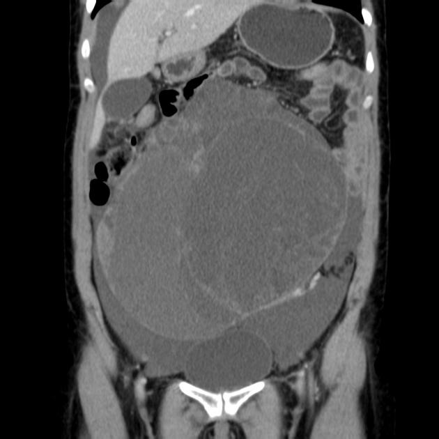
In general, the cell type (e.g. serous, mucinous) often cannot be determined by the appearance on imaging 5. While some of the specific features can vary between the subtypes, there are certain features which are more common among mucinous tumors: 3
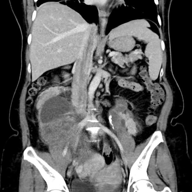
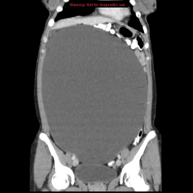
- often larger than their serous counterparts (on occasion they may be enormous)
- tend to be more multilocular with small cystic components +/- honeycomb-like locules
- calcification is comparatively rare and if present tends to be linear
- usually unilateral
- peritoneal carcinomatosis is less common compared with serous tumors
- may have accompanying pseudomyxoma peritonei
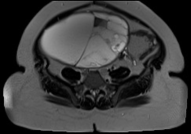
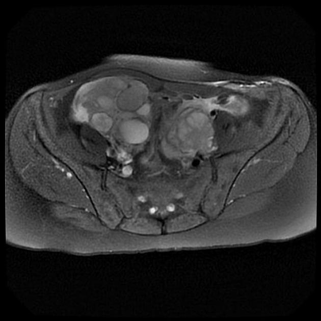
MRI
-
T1
- signal intensity of locules varies depending on the degree of mucin concentration
- loculi with watery mucin have a lower signal intensity than loculi with thicker mucin
-
T2
- the above-mentioned corresponding signal intensities are flipped so that loculi with watery mucin have a higher signal intensity and loculi with thicker mucin appear slightly hypointense
- the combination of locules with different signal intensities may result in a "stained glass appearance"
Treatment and prognosis
Prognosis of mucinous tumors is highly dependent on the stage and histologic composition: see individual subtypes for further details. Primary treatment is surgical unless there is extra-ovarian disease 6.
Differential diagnosis
-
ovarian serous tumors
- tend to be unilocular
- often smaller than mucinous tumors
- more frequently bilateral
- calcifications (psammoma calcifications)
-
hemorrhagic cyst
- smaller
- unilocular
- resolves on a follow-up scan
-
endometrioma
- high signal on T1 weighted images with T2 shading (signal characteristics typical of blood products)


 Unable to process the form. Check for errors and try again.