Phrenic nerve paralysis (paresis or palsy) has many causes and can be caused by lesions anywhere along the course of the phrenic nerve from the neck to the medial diaphragm adjacent to the pericardium.
On this page:
Epidemiology
The epidemiology depends on the underlying cause, see below.
Clinical presentation
Presentation will also depend on the underlying cause and comorbidities. Most cases are unilateral and asymptomatic 1,7. Dyspnea is exacerbated by lying flat (orthopnea) or immersion in water and can be severe when both hemidiaphragms are weak or paralyzed. Respiratory function tests show reduced total lung capacity and a restrictive pattern 1.
Phrenic nerve conduction studies can be carried out with placement of an esophageal electrode to record diaphragmatic contractions and stimulation of the nerve in the neck either with surface stimulation or with a monopolar needle electrode at the level of the cricoid cartilage 7. Alternatively, diaphragmatic electromyography may be carried out. The two tests are complementary 7.
Pathology
Etiology
Many cases are idiopathic, possibly postviral, and may recover spontaneously 8,9. Common known causes include:
-
malignancy
mediastinal tumors or neck malignancy
-
trauma and iatrogenic
penetrating injury
chiropractic manipulation 1
postoperative: especially cardiac: up to 10% of cases 7,9,11
-
direct trauma
compression by hematoma
local anesthetic infiltration
-
intrascalene brachial plexus nerve blocks
incidence reduced with ultrasound guidance and use of lower volumes of local anesthetic 12
forceps delivery (newborn) 5
-
neuromuscular diseases
-
inflammation
-
direct compression
cervical osteophytes
Radiographic features
Plain radiograph
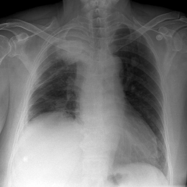
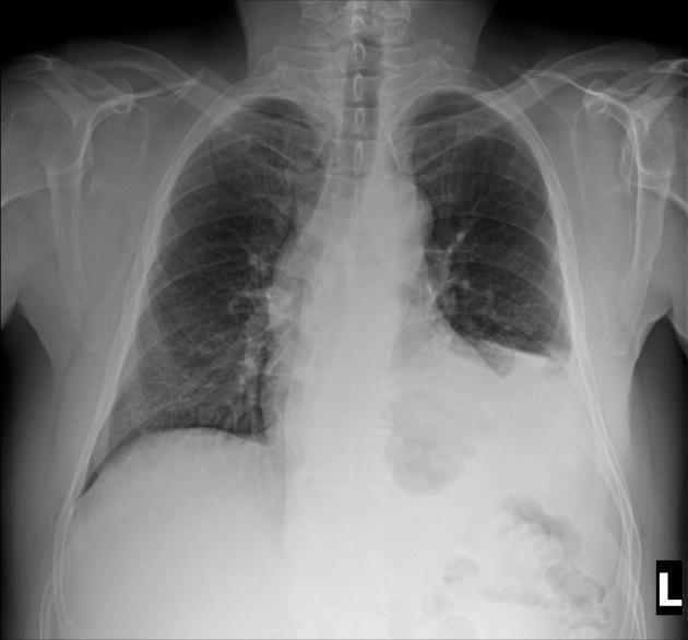
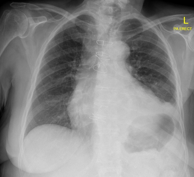
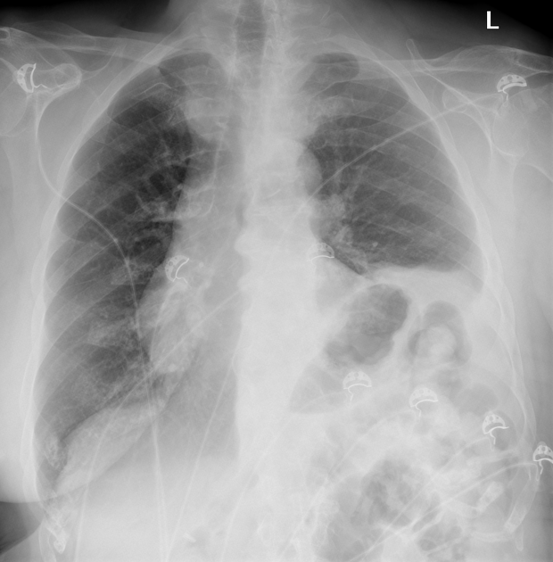
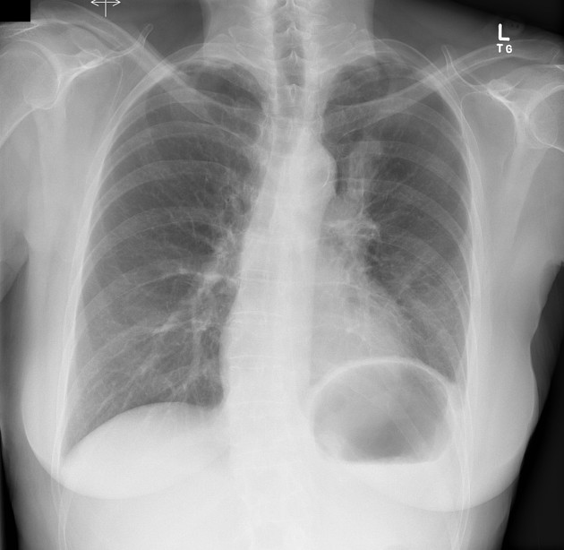
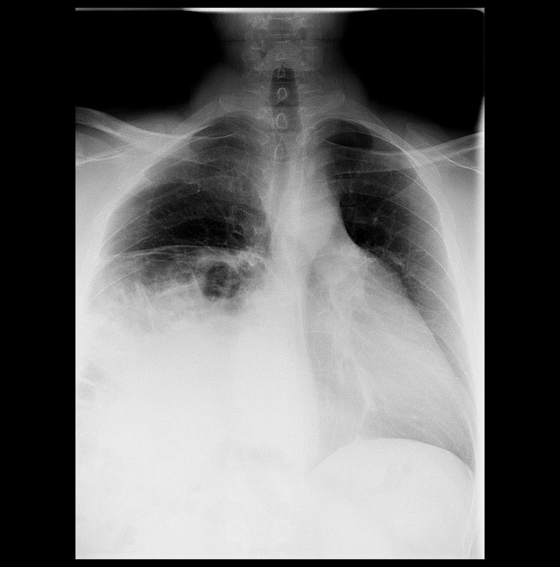
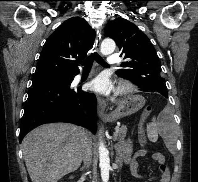
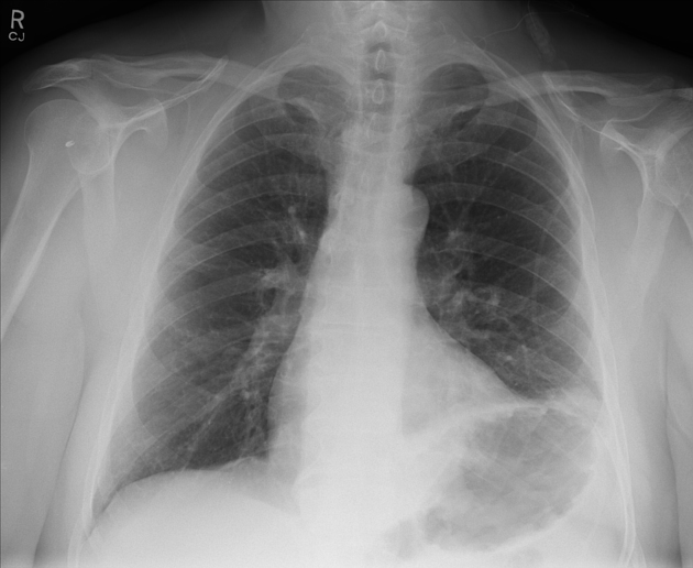
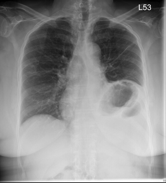
In some cases the diagnosis is obvious. However, as diaphragmatic position is asymmetric, an understanding of the normal level of the diaphragms is important (see normal position of diaphragms on chest radiography). If the left hemidiaphragm is higher than the right or the right is higher than the left by more than ~2 centimeters, one of the many causes of diaphragmatic elevation should be sought. This, of course, includes phrenic nerve paralysis 10.
Fluoroscopy
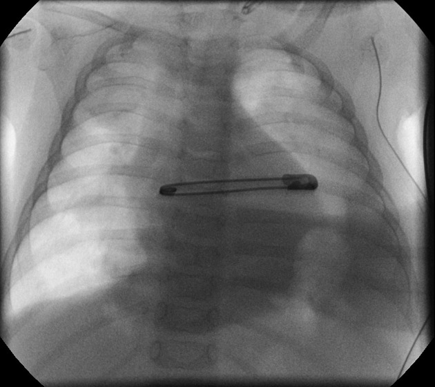
In normal subjects, both hemidiaphragms descend with inspiration or sniffing. In cases of phrenic nerve paralysis, the affected side demonstrates paradoxical upward movement 10.
Interpretation of diaphragmatic fluoroscopy can be difficult and this is due partly to normal variable contribution of thoracic, diaphragmatic and abdominal contributions to ventilation which can be minimized by supine positioning, see sniff test.
Fluoroscopy may also be employed at the time of nerve conduction studies to confirm diaphragmatic movement.
Ultrasound
Ultrasound allows for evaluation of diaphragmatic movements, and may also be employed at the time of nerve conduction studies to confirm needle placement.
CT
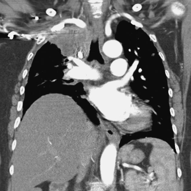
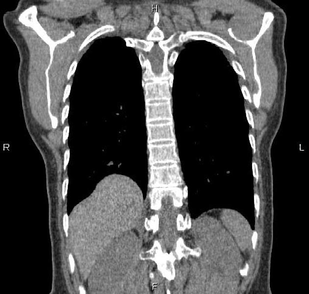
CT may show atrophy of the diaphragmatic crus and may identify the cause of the phrenic nerve palsy.
MRI
MRI is particularly suited to evaluation of superior sulcus tumors, and is better able to determine the extent of tumor in such cases.
Treatment and prognosis
In permanent symptomatic unilateral or bilateral paralysis diaphragmatic pacing may be used. This can either take the form of distal phrenic nerve stimulation or direct muscular stimulation with implanted electrodes 8.
Prognosis is influenced by the underlying cause. Nerve function does not usually recover in cases of neoplastic involvement. Like other compressive neuropathies gradual improvement may take place over many months or years 1.
Differential diagnosis
The differential of a phrenic nerve paralysis is essentially that of an elevated hemidiaphragm with common entities to be considered being:
-
masses/collections pushing the diaphragm up from below
hepatic mass
intra-abdominal fat
-
reduced volume of the lung pulling the diaphragm up
In cases of bilateral diaphragmatic paralysis, in addition to bilateral phrenic nerve palsy a number of conditions should be considered 8:
high spinal cord injury
shrinking lung syndrome in autoimmune disease, especially systemic lupus erythematosus





 Unable to process the form. Check for errors and try again.