Citation, DOI, disclosures and article data
Citation:
Weerakkody Y, Murphy A, Lukies M, et al. Septic pulmonary emboli. Reference article, Radiopaedia.org (Accessed on 06 Jun 2023) https://doi.org/10.53347/rID-22848
Septic pulmonary emboli refer to the embolization of infectious particles (intravascular thrombus containing microorganisms) into the lungs via the pulmonary arterial system.
Symptoms can be not specific but most manifest as a bacteremia 18 with, dyspnea, chest pain, cough and other respiratory symptoms. Often concurrent symptoms of the extrapulmonary primary infective focus are also present.
Septic emboli can originate from different sources 5:
- right-sided infective endocarditis, particularly tricuspid valve (occasionally pulmonary valve 19)
- infection elsewhere in the body (e.g. soft tissue infection) with associated septal defects
- infected deep venous thrombosis
- immunological deficiencies
- infected catheters/lines
- post-anginal septicemia 10
- periodontal disease 10
Common pathogens include 22:
- Klebsiella pneumoniae
- Staphylococcus aureus
- Pseudomonas aeruginosa
- Escherichia coli
-
Salmonella group B
-
Fusobacterium necrophorum (in Lemierre syndrome)
The clinical context is significant in image interpretation and differential considerations. Most patients will be clinically septic and have positive blood cultures at the time of imaging assessment.
Plain radiograph
Chest x-ray features are nonspecific but may show 3:
- peripheral, lower lobe predominant infiltrative densities: can be unilateral or bilateral
- diffuse bilateral nodular densities (often poorly marginated) in varying stages of cavitation
- these nodules generally range between 1-3 cm
- the nodules vary greatly in size, which is a reflection of repeated episodes of embolic shower 15
- may increase in number or change in appearance (size or degree of cavitation) on subsequent short-term follow up radiographs 12
- accompanying small pleural effusions can be common
- features suggestive of complicating pleura empyemas may be seen
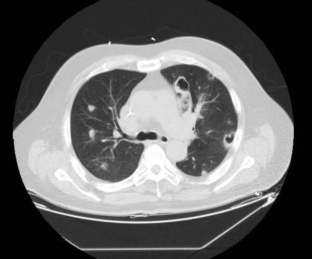
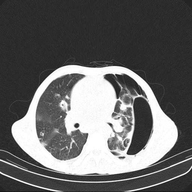
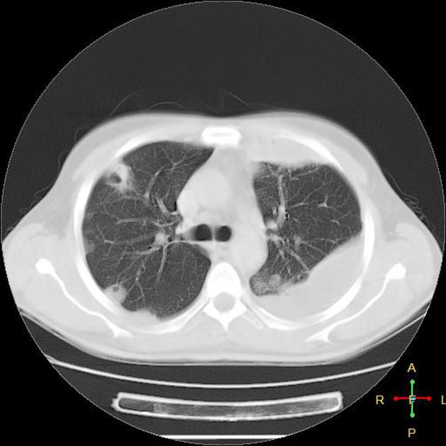
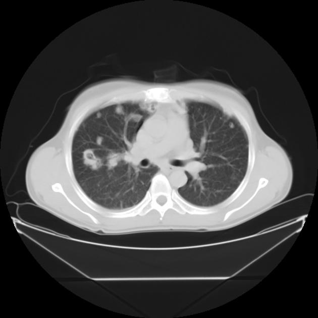
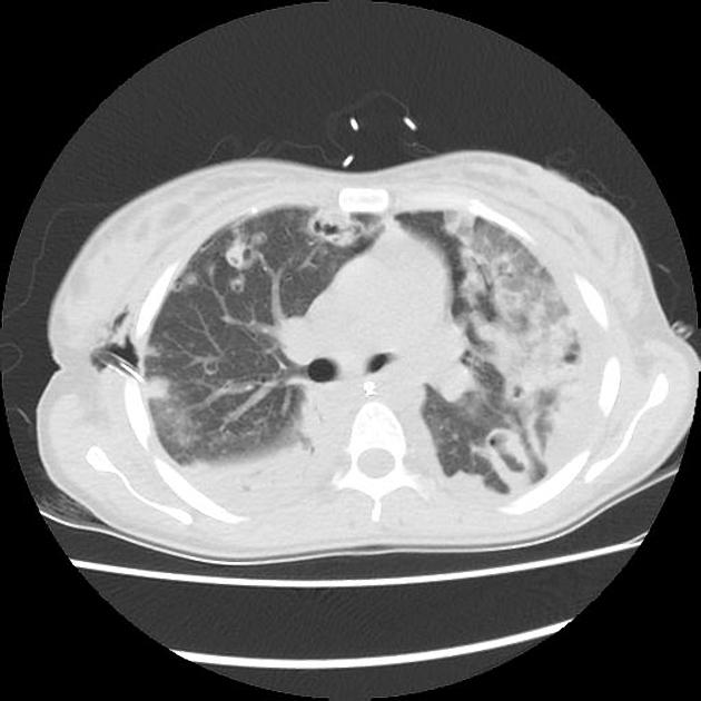
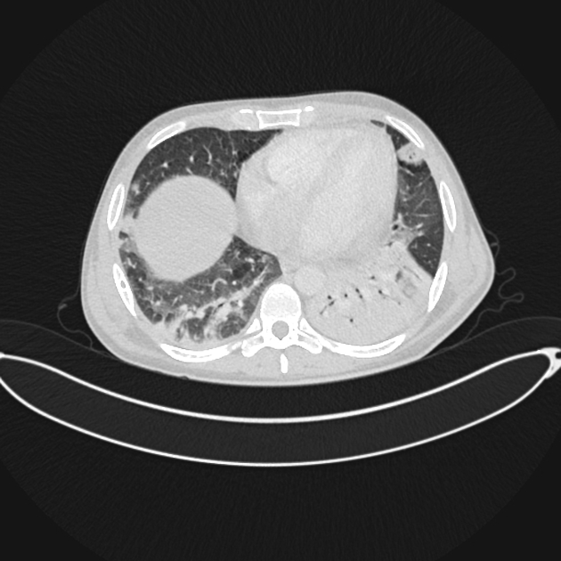
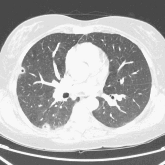
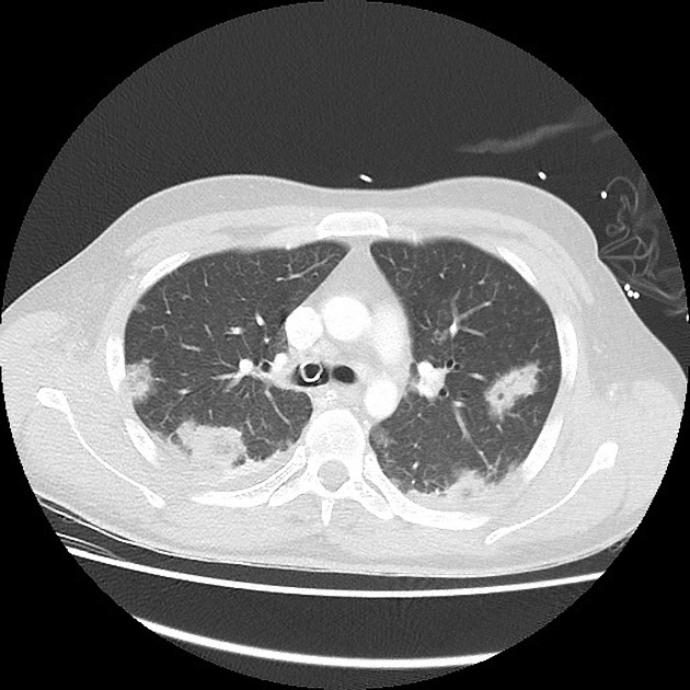
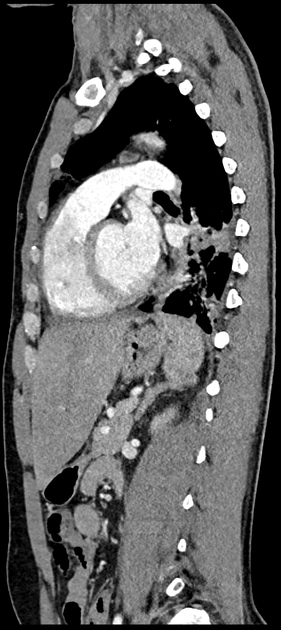
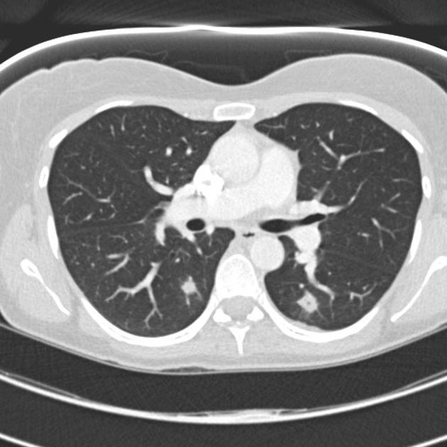
CT
Bilateral abnormalities may be present in as much as 80% of cases 18.
-
feeding vessel sign 9: peripheral nodules with clearly identifiable feeding vessels associated with lung abscesses 2,3
- subpleural nodular lesions or wedge-shaped densities with or without necrosis caused by septic infarcts (these can manifest as cavitary pulmonary infarcts)
- these may have a dependent, lower zone predication 13,15
- the wedge-shaped lesions usually range between 10-20 mm 6
- peripheral nodular densities usually range between 5-35 mm 7
Treatment and prognosis
Management focuses on treatment of the underlying infection and any associated complications.
Complications
Recognized complications include:
For small and/or different size cavitary lung lesions on HRCT or CT chest, consider:
- 1. Jaffe RB, Koschmann EB. Septic pulmonary emboli. Radiology. 1970;96 (3): 527-32. Radiology (abstract) - doi:10.1148/96.3.527 - Pubmed citation
- 2. Wong KS, Lin TY, Huang YC et-al. Clinical and radiographic spectrum of septic pulmonary embolism. Arch. Dis. Child. 2002;87 (4): 312-5. Arch. Dis. Child. (full text) - doi:10.1136/adc.87.4.312 - Free text at pubmed - Pubmed citation
- 3. Huang RM, Naidich DP, Lubat E et-al. Septic pulmonary emboli: CT-radiographic correlation. AJR Am J Roentgenol. 1989;153 (1): 41-5. doi:10.2214/ajr.153.1.41 - Pubmed citation
- 4. Zelefsky MN, Lutzker LG. The target sign: a new radiologic sign of septic pulmonary emboli. AJR Am J Roentgenol. 1977;129 (3): 453-5. doi:10.2214/ajr.129.3.453 - Pubmed citation
- 5. Engelke C, Schaefer-Prokop C, Schirg E et-al. High-resolution CT and CT angiography of peripheral pulmonary vascular disorders. Radiographics. 2002;22 (4): 739-64. Radiographics (full text) - Pubmed citation
- 6. Iwasaki Y, Nagata K, Nakanishi M et-al. Spiral CT findings in septic pulmonary emboli. Eur J Radiol. 2001;37 (3): 190-4. Pubmed citation
- 7. Kuhlman JE, Fishman EK, Teigen C. Pulmonary septic emboli: diagnosis with CT. Radiology. 1990;174 (1): 211-3. Radiology (abstract) - Pubmed citation
- 8. Kwon WJ, Jeong YJ, Kim KI et-al. Computed tomographic features of pulmonary septic emboli: comparison of causative microorganisms. J Comput Assist Tomogr. 2007;31 (3): 390-4. doi:10.1097/01.rct.0000243455.23308.a9 - Pubmed citation
- 9. Dodd JD, Souza CA, MüLler NL. High-resolution MDCT of pulmonary septic embolism: evaluation of the feeding vessel sign. AJR Am J Roentgenol. 2006;187 (3): 623-9. doi:10.2214/AJR.05.0681 - Pubmed citation
- 10. Mattar CS, Keith RL, Byrd RP et-al. Septic pulmonary emboli due to periodontal disease. Respir Med. 2006;100 (8): 1470-4. doi:10.1016/j.rmed.2005.11.010 - Pubmed citation
- 11. Cervia JS, Caputo TA, Davis SD et-al. Septic pulmonary embolism complicating a central venous catheter. Chest. 01;98 (6): 1526. doi:10.1378/chest.98.6.1526 - Pubmed citation
- 12. Rossi SE, Goodman PC, Franquet T. Nonthrombotic pulmonary emboli. AJR Am J Roentgenol. 2000;174 (6): 1499-508. doi:10.2214/ajr.174.6.1741499 - Pubmed citation
- 13. Khashper A, Discepola F, Kosiuk J et-al. Nonthrombotic pulmonary embolism. AJR Am J Roentgenol. 2012;198 (2): W152-9. doi:10.2214/AJR.11.6407 - Pubmed citation
- 14. Jorens PG, Van Marck E, Snoeckx A et-al. Nonthrombotic pulmonary embolism. Eur. Respir. J. 2009;34 (2): 452-74. Eur. Respir. J. (full text) - doi:10.1183/09031936.00141708 - Pubmed citation
- 15. Han D, Lee KS, Franquet T et-al. Thrombotic and nonthrombotic pulmonary arterial embolism: spectrum of imaging findings. Radiographics. 2003;23 (6): 1521-39. Radiographics (full text) - doi:10.1148/rg.1103035043 - Pubmed citation
- 16. Sheu CC, Hwang JJ, Tsai JR et-al. Spontaneous pneumothorax as a complication of septic pulmonary embolism in an intravenous drug user: a case report. Kaohsiung J. Med. Sci. 2006;22 (2): 89-93. doi:10.1016/S1607-551X(09)70226-8 - Pubmed citation
- 17. Corzo JE, Lozano De LeóN F, GóMez-Mateos J et-al. Pneumothorax secondary to septic pulmonary emboli in tricuspid endocarditis. Thorax. 1993;47 (12): 1080-1. Free text at pubmed - Pubmed citation
- 18. Ye R, Zhao L, Wang C, Wu X, Yan H. Clinical characteristics of septic pulmonary embolism in adults: a systematic review. (2014) Respiratory medicine. 108 (1): 1-8. doi:10.1016/j.rmed.2013.10.012 - Pubmed
- 19. Inchaustegui CA, Wang KY, Teniola O, De Rosen VL. Large septic pulmonary embolus complicating streptococcus mutans pulmonary valve endocarditis. (2018) Journal of radiology case reports. 12 (2): 18-27. doi:10.3941/jrcr.v12i2.3240 - Pubmed
- 20. Ho VT, Rothenberg KA, McFarland G, Tran K, Aalami OO. Septic Pulmonary Emboli From Peripheral Suppurative Thrombophlebitis: A Case Report and Literature Review. (2018) Vascular and endovascular surgery. 52 (8): 633-635. doi:10.1177/1538574418779469 - Pubmed
- 21. Baidya A, Ganakumar V, Jadon RS, Ranjan P, Manchanda S, Sood R. Septic pulmonary emboli as a complication of peripheral venous cannula insertion. (2018) Drug discoveries & therapeutics. 12 (2): 111-113. doi:10.5582/ddt.2018.01016 - Pubmed
- 22. Chou DW, Wu SL, Chung KM, Han SC, Cheung BM. Septic Pulmonary Embolism Requiring Critical Care: Clinicoradiological Spectrum, Causative Pathogens and Outcomes. (2016) Clinics (Sao Paulo, Brazil). 71 (10): 562-569. doi:10.6061/clinics/2016(10)02 - Pubmed
Promoted articles (advertising)