
Tubal ectopic pregnancy, also known as adnexal ectopic pregnancy, is the most common location of an ectopic pregnancy.
On this page:
Epidemiology
It is the most common type of ectopic by far, accounting for ~95% (range 93-97%) of cases.
Diagnosis
On transvaginal imaging, findings indicating a definitive diagnosis of a tubal ectopic pregnancy include:
extra-ovarian adnexal mass comprising a gestational sac containing a yolk sac
extra-ovarian adnexal mass comprising a gestational sac and fetal pole (with or without cardiac activity)
On transvaginal imaging, findings indicating a high probability of a tubal ectopic pregnancy include:
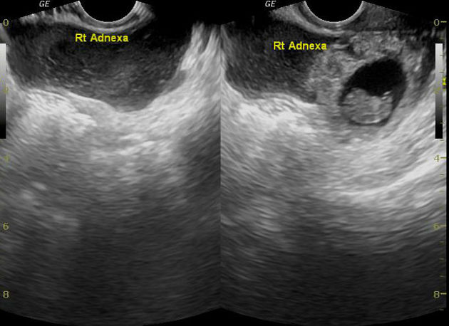
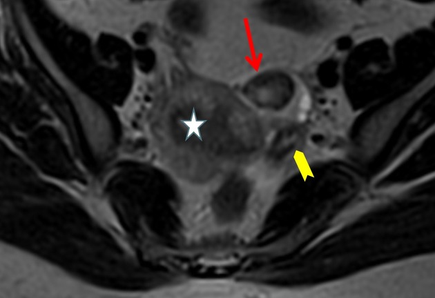
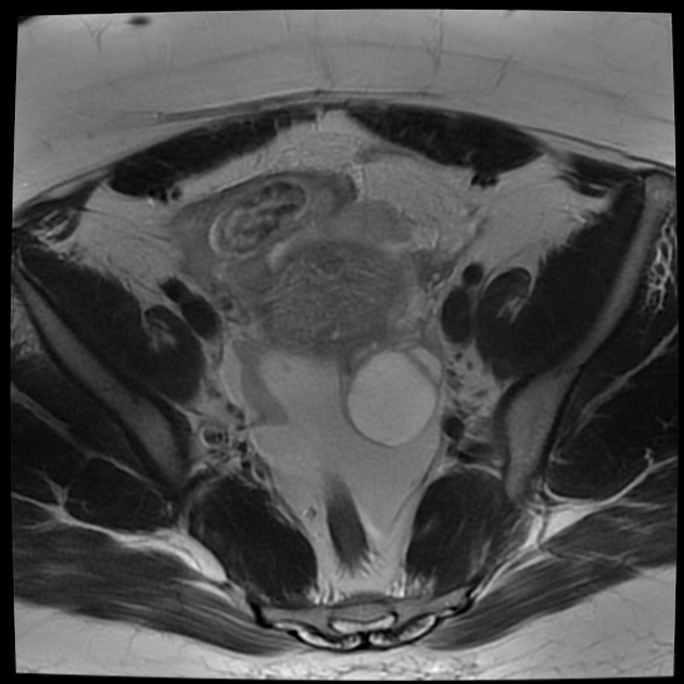
extra-ovarian adnexal mass with an empty gestational sac (tubal ring sign or bagel sign)
extra-ovarian heterogeneous adnexal mass (blob sign)
On transvaginal imaging, findings indicating possible tubal ectopic pregnancy include:
empty uterus
fluid within the uterine cavity (pseudosac)
The clinical presentation, serum hCG levels, uterine, and adnexal findings must be considered together.
Pathology
Although the fallopian tube has many anatomical parts, for the purposes of ectopic location, it can be divided into 1:
-
considered the longest segment
most common site, accounting for ~70%
isthmic ectopic: ~12%
fimbrial ectopic: ~11%
Radiographic features
Ultrasound
-
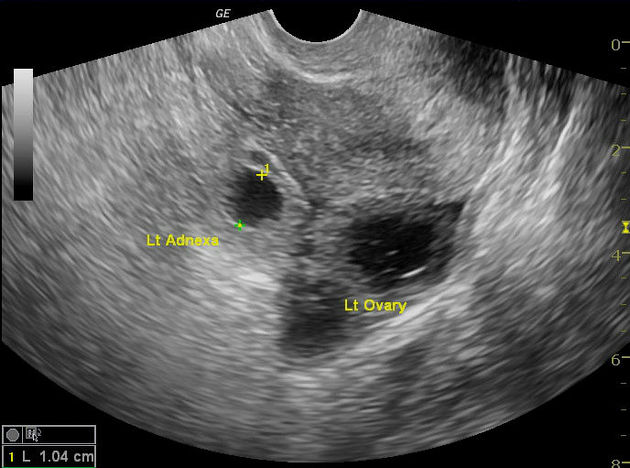
an adnexal mass that is separate from the ovary is the most common finding and may be seen in ~95% (range 89-100%) of cases 1
the presence of an adnexal mass becomes more specific for an ectopic pregnancy when it contains a yolk sac or a living embryo or when it moves independently from the ovary
an extrauterine mass may not be sonographically detected in up to 35% of patients with an ectopic pregnancy
usually, the corpus luteum is on the same side as ectopic gestational sac 2; rupture of the cyst can present with abdominal pain and hemoperitoneum mimicking ectopic rupture
there may be evidence of a hematosalpinx (a tubal ectopic is the commonest cause for a hematosalpinx 3)
-
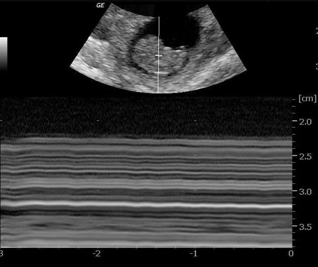
tubal echogenic ring
typically a 1-3 cm mass consisting of a 2-4 mm concentric, echogenic rim of tissue surrounding a hypoechoic center
represents an echogenic ring surrounding an extrauterine gestational sac
-
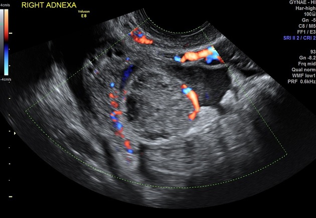
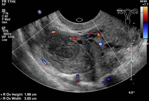
color Doppler interrogation may show peripheral vascularity giving a ring of fire sign
a corpus luteum may have similar color Doppler flow
large hemoperitoneum or large amount of free fluid, especially in cases of rupture, may be present
Treatment and prognosis
Compared to an interstitial ectopic or cervical ectopic, the risk of uncontrollable hemorrhage is fortunately lower. Nonetheless, expedient diagnosis is required.
Management was previously only surgical, with open and then laparoscopic salpingectomy being favored. Salpingotomy (thus preserving the tube) is increasingly performed.
Medical management includes methotrexate (a folate antagonist) either administered systemically or by direct ultrasound-guided injection. After methotrexate therapy, the ectopic pregnancy may show a paradoxical increase in size and vascularity on subsequent imaging even with successful methotrexate.
Increasingly, conservative management is being recognized as an option for ectopic pregnancy where rupture has not occurred (i.e. no hemoperitoneum) and fetal demise has already taken place.
Complications
Differential diagnosis
other types of ectopic pregnancy 4
-
may also present as an "adnexal mass" in a patient in whom there is clinical suspicion for ectopic pregnancy and may have similar color Doppler flow
should be attached to the ovary, whereas a tubal ectopic will slide separately from the ovary with transducer pressure


 Unable to process the form. Check for errors and try again.