
Bone scintigraphy
Updates to Article Attributes
Body
was changed:
Bone scanscans isare a nuclear medicine (scintigraphic) study that makes use of Technetium 99m (commonly Tc99m-methylene diphosphonate (MDP)) as the active agent.
The study has three stagesphases which follow intravenous injection of the tracer. Sometimes a fourth (delayed/delayed) phase is performed.
Clinical indications
- malignancy: detection and follow-up of skeletal metastases
- detection of radiographically occult fractures, e.g. stress or insufficiency fractures
- osteomyelitis
- reflex sympathetic dystrophy
- hip joint prosthesis: evaluation for infection or loosening
Patient preparation
- hydration
- remove metal objects
- void immediately before study
Tracer dose and route of administration
Tc99m diphosphonate is administered intravenously, at a dose of 20 mCi.
The phasesPhases
Flow phase
- 2
to 5-sec-to-5 second images are obtained for 60 seconds after injection - demonstrates perfusion and
characterisescharacterises the blood flow to a particular area
Blood pool phase
- the
bloodblood-pool image is obtained 5minminutes after injection - demonstrates the blood pool, not the blood flow
- inflammation causes capillary dilatation and increased blood flow
If the study is going to be a triphasic bone scan, a third phase is added.
Delayed phase
- the bone image
isis obtained 2-4 hours later - urinary excretion has decreased the
amountamount of the radionuclide in soft tissue - mechanism of uptake is not uncertainly known
- degree of uptake depends on blood flow and rate of new bone formation
Delayed/delayed
- obtained 24 hours after injection as a static image
See also
-<p><strong>Bone scan</strong> is a nuclear medicine (scintigraphic) study that makes use of <a href="/articles/technetium-99m">Technetium <sup>99</sup>m</a> (commonly Tc<sup>99</sup>m-methylene diphosphonate (MDP)) as the active agent.</p><p>The study has three stages which follow intravenous injection of the tracer.</p><h4>Clinical indications</h4><ul>- +<p><strong>Bone scans</strong> are a nuclear medicine (scintigraphic) study that makes use of <a href="/articles/technetium-99m">Technetium <sup>99</sup>m</a> (commonly Tc<sup>99</sup>m-methylene diphosphonate (MDP)) as the active agent.</p><p>The study has three phases which follow intravenous injection of the tracer. Sometimes a fourth (delayed/delayed) phase is performed. </p><h4>Clinical indications</h4><ul>
-<li>detection of radiographically occult fractures</li>- +<li>detection of radiographically occult fractures, e.g. <a title="Stress fracture" href="/articles/stress-fractures">stress</a> or <a title="Insufficiency fractures" href="/articles/insufficiency-fracture">insufficiency fractures</a>
- +</li>
-</ul><h4>Tracer dose and route of administration</h4><p>Tc99m diphosphonate is administered intravenously, at a dose of 20 mCi.</p><h4>The phases</h4><h5>Flow phase</h5><ul>-<li>2 to 5-sec images are obtained for 60 seconds after injection</li>-<li>demonstrates perfusion and characterises the blood flow to a particular area</li>- +</ul><h4>Tracer dose and route of administration</h4><p>Tc99m diphosphonate is administered intravenously, at a dose of 20 mCi.</p><h4>Phases</h4><h5>Flow phase</h5><ul>
- +<li>2-to-5 second images are obtained for 60 seconds after injection</li>
- +<li>demonstrates perfusion and characterises the blood flow to a particular area</li>
-<li>the blood-pool image is obtained 5 min after injection</li>- +<li>the blood-pool image is obtained 5 minutes after injection</li>
-<li>the bone image is obtained 2-4 hours later</li>-<li>urinary excretion has decreased the amount of the radionuclide in soft tissue</li>-</ul><h4>See also</h4><ul>- +<li>the bone image is obtained 2-4 hours later</li>
- +<li>urinary excretion has decreased the amount of the radionuclide in soft tissue</li>
- +<li>mechanism of uptake is not uncertainly known</li>
- +<li>degree of uptake depends on blood flow and rate of new bone formation</li>
- +</ul><h5>Delayed/delayed</h5><ul><li>obtained 24 hours after injection as a static image</li></ul><h4>See also</h4><ul>
References changed:
- 2. Love C, Din A, Tomas M, Kalapparambath T, Palestro C. Radionuclide Bone Imaging: An Illustrative Review. Radiographics. 2003;23(2):341-58. <a href="https://doi.org/10.1148/rg.232025103">doi:10.1148/rg.232025103</a> - <a href="https://www.ncbi.nlm.nih.gov/pubmed/12640151">Pubmed</a>
- 3. Alazraki N, Dries D, Datz F, Lawrence P, Greenberg E, Taylor A. Value of a 24-Hour Image (Four-Phase Bone Scan) in Assessing Osteomyelitis in Patients with Peripheral Vascular Disease. J Nucl Med. 1985;26(7):711-7. - <a href="https://www.ncbi.nlm.nih.gov/pubmed/3159858">Pubmed</a>
Images Changes:
Image 1 Nuclear medicine ( update )
Caption
was changed:
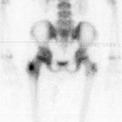
Case 1: Bone scan showing osteoid osteoma
Image 4 Nuclear medicine ( create )


 Unable to process the form. Check for errors and try again.