Calcifying fibrous tumor
Updates to Article Attributes
Calcifying fibrous tumorstumours, previously known as calcifying fibrous pseudotumorspseudotumours, are rare, benign fibroblastic tumorstumours of the soft tissues.
Epidemiology
It can occur at all ages and there is no strong gender predilection 1. Fewer than 200 cases have been reported in the English literature 1.
Clinical presentation
Most cases are asymptomatic, so the tumortumour tends to be found incidentally on imaging 1. When symptoms are present, they are non-specific and include a palpable painless mass or localizedlocalised pain 1.
Pathology
Location
Calcifying fibrous tumorstumours can occur throughout the body's subcutaneous or deep soft tissues, with a few sites being more commonly reported 1:
- stomach (18%) 6
- pleura (10%)
- small intestine (9%) 5
- peritoneum (7%) 3
- neck (6%) 8
- mesentery (5%)
Many other sites have been described, including calcifying fibrous tumortumour of the lung, liver 2, and between muscles 8. Association with bone is unusual but a case involving the clivus has been reported 4.
Classification
The entity is included in the WHO classification of tumorstumours of soft tissue under the fibroblastic/myofibroblastic tumorstumours category.
Macroscopic appearance
It is a circumscribed, nonencapsulated mass and occasionally infiltrates surrounding tissues 1.
Microscopic appearance
The lesion is composed predominantly of hyalinized stroma with interspersed psammomatous and dystrophic calcifications, fibroblastic spindle cells, and mononuclear/lymphoplasmacytic inflammatory infiltrate 1,3.
Radiographic features
The appearance depends on the site but in general reflect the histology 1-7.
Plain radiograph
A mass is present with calcifications.
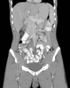
CT
Partially calcified mass is demonstrated with some contrast enhancement, which progresses on delayed images.
Ultrasound
The mass is hypoechoic with acoustic shadowing and foci of hyperechoic calcifications.
MRI
The mass has the following signal characteristics suggesting fibrosis/collagenous content:
- T1: slightly low signal intensity
- T2: low signal intensity
- T1 C+: relatively hypovascular but with slow progressive enhancement
Treatment and prognosis
Surgical excision is curative. The condition is benign, without reports of mortality; recurrence is rare 1.
Differential diagnosis
The differential diagnosis is broad and depends on location but includes mesenchymal tumorstumours such as 1:
-<p><strong>Calcifying fibrous tumors</strong>, previously known as <strong>calcifying fibrous pseudotumors</strong>, are rare, benign fibroblastic tumors of the soft tissues.</p><h4>Epidemiology</h4><p>It can occur at all ages and there is no strong gender predilection <sup>1</sup>. Fewer than 200 cases have been reported in the English literature <sup>1</sup>.</p><h4>Clinical presentation</h4><p>Most cases are asymptomatic, so the tumor tends to be found incidentally on imaging <sup>1</sup>. When symptoms are present, they are non-specific and include a palpable painless mass or localized pain <sup>1</sup>.</p><h4>Pathology</h4><h5>Location</h5><p>Calcifying fibrous tumors can occur throughout the body's subcutaneous or deep soft tissues, with a few sites being more commonly reported <sup>1</sup>:</p><ul>- +<p><strong>Calcifying fibrous tumours</strong>, previously known as <strong>calcifying fibrous pseudotumours</strong>, are rare, benign fibroblastic tumours of the soft tissues.</p><h4>Epidemiology</h4><p>It can occur at all ages and there is no strong gender predilection <sup>1</sup>. Fewer than 200 cases have been reported in the English literature <sup>1</sup>.</p><h4>Clinical presentation</h4><p>Most cases are asymptomatic, so the tumour tends to be found incidentally on imaging <sup>1</sup>. When symptoms are present, they are non-specific and include a palpable painless mass or localised pain <sup>1</sup>.</p><h4>Pathology</h4><h5>Location</h5><p>Calcifying fibrous tumours can occur throughout the body's subcutaneous or deep soft tissues, with a few sites being more commonly reported <sup>1</sup>:</p><ul>
-</ul><p>Many other sites have been described, including <a href="/articles/calcifying-fibrous-pseudotumour-of-the-lung">calcifying fibrous tumor of the lung</a>, liver <sup>2</sup>, and between muscles <sup>8</sup>. Association with bone is unusual but a case involving the clivus has been reported <sup>4</sup>.</p><h5>Classification</h5><p>The entity is included in the <a href="/articles/who-classification-of-tumors-of-soft-tissue">WHO classification of tumors of soft tissue</a> under the fibroblastic/myofibroblastic tumors category.</p><h5>Macroscopic appearance</h5><p>It is a circumscribed, nonencapsulated mass and occasionally infiltrates surrounding tissues <sup>1</sup>.</p><h5>Microscopic appearance</h5><p>The lesion is composed predominantly of hyalinized stroma with interspersed psammomatous and dystrophic calcifications, fibroblastic spindle cells, and mononuclear/lymphoplasmacytic inflammatory infiltrate <sup>1,3</sup>.</p><h4>Radiographic features</h4><p>The appearance depends on the site but in general reflect the histology <sup>1-7</sup>.</p><h5>Plain radiograph</h5><p>A mass is present with calcifications.</p><h5>CT</h5><p>Partially calcified mass is demonstrated with some contrast enhancement, which progresses on delayed images.</p><h5>Ultrasound</h5><p>The mass is hypoechoic with acoustic shadowing and foci of hyperechoic calcifications.</p><h5>MRI</h5><p>The mass has the following signal characteristics suggesting fibrosis/collagenous content:</p><ul>- +</ul><p>Many other sites have been described, including <a href="/articles/calcifying-fibrous-pseudotumour-of-the-lung">calcifying fibrous tumour of the lung</a>, liver <sup>2</sup>, and between muscles <sup>8</sup>. Association with bone is unusual but a case involving the clivus has been reported <sup>4</sup>.</p><h5>Classification</h5><p>The entity is included in the <a href="/articles/who-classification-of-tumors-of-soft-tissue">WHO classification of tumours of soft tissue</a> under the fibroblastic/myofibroblastic tumours category.</p><h5>Macroscopic appearance</h5><p>It is a circumscribed, nonencapsulated mass and occasionally infiltrates surrounding tissues <sup>1</sup>.</p><h5>Microscopic appearance</h5><p>The lesion is composed predominantly of hyalinized stroma with interspersed psammomatous and dystrophic calcifications, fibroblastic spindle cells, and mononuclear/lymphoplasmacytic inflammatory infiltrate <sup>1,3</sup>.</p><h4>Radiographic features</h4><p>The appearance depends on the site but in general reflect the histology <sup>1-7</sup>.</p><h5>Plain radiograph</h5><p>A mass is present with calcifications.</p><h5>CT</h5><p>Partially calcified mass is demonstrated with some contrast enhancement, which progresses on delayed images.</p><h5>Ultrasound</h5><p>The mass is hypoechoic with acoustic shadowing and foci of hyperechoic calcifications.</p><h5>MRI</h5><p>The mass has the following signal characteristics suggesting fibrosis/collagenous content:</p><ul>
-</ul><h4>Treatment and prognosis</h4><p>Surgical excision is curative. The condition is benign, without reports of mortality; recurrence is rare <sup>1</sup>.</p><h4>Differential diagnosis</h4><p>The differential diagnosis is broad and depends on location but includes mesenchymal tumors such as <sup>1</sup>:</p><ul>-<li><a href="/articles/inflammatory-myofibroblastic-tumour">inflammatory myofibroblastic tumor</a></li>-<li><a href="/articles/solitary-fibrous-tumour">solitary fibrous tumor</a></li>-<li><a href="/articles/gastrointestinal-stromal-tumour-1">gastrointestinal stromal tumor</a></li>- +</ul><h4>Treatment and prognosis</h4><p>Surgical excision is curative. The condition is benign, without reports of mortality; recurrence is rare <sup>1</sup>.</p><h4>Differential diagnosis</h4><p>The differential diagnosis is broad and depends on location but includes mesenchymal tumours such as <sup>1</sup>:</p><ul>
- +<li><a href="/articles/inflammatory-myofibroblastic-tumour">inflammatory myofibroblastic tumour</a></li>
- +<li><a href="/articles/solitary-fibrous-tumour">solitary fibrous tumour</a></li>
- +<li><a href="/articles/gastrointestinal-stromal-tumour-1">gastrointestinal stromal tumour</a></li>
References changed:
- 9. Turbiville D & Zhang X. Calcifying Fibrous Tumor of the Gastrointestinal Tract: A Clinicopathologic Review and Update. WJG. 2020;26(37):5597-605. <a href="https://doi.org/10.3748/wjg.v26.i37.5597">doi:10.3748/wjg.v26.i37.5597</a> - <a href="https://www.ncbi.nlm.nih.gov/pubmed/33071524">Pubmed</a>
- 10. Sabrine D, Hafsa E, Amine R, Zakia B, Fouad Z. Calcifying Fibrous Tumor of the Mesentery: A Case Report and a Review of the Literature. Clin Med Insights Pathol. 2020;13:2632010X2093068. <a href="https://doi.org/10.1177/2632010x20930689">doi:10.1177/2632010x20930689</a> - <a href="https://www.ncbi.nlm.nih.gov/pubmed/32637936">Pubmed</a>
Image 1 CT (C+ portal venous phase) ( create )


 Unable to process the form. Check for errors and try again.