
Forearm series (pediatric)
Updates to Article Attributes
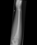
The forearm series for paediatrics comprises an anteroposterior and lateral projection. These projections examine the entire radius and ulna including the distal and proximal articulations.
Indications
Forearm x-rays are indicated for a variety of settings including:
- trauma
- bony tenderness
- suspected fracture
- obvious deformity
- suspected foreign body
- infection
Projections
Standard projections
-
anteroposterior view
- demonstrates radius and ulna in natural anatomical position
- lateral view
Modified trauma projections
Where patients are in a considerable amount of pain, these two projections may replace the standard projections instead to obtain diagnostic images of the radius and ulna whilst requiring little to no patient movement.
-
posteroanterior view
- demonstrates PA wrist distally and lateral elbow proximally
-
horizontal beam lateral view
- demonstrates lateral wrist distally and AP elbow proximally
Patient preparation
Patients should remove any jewellery or clothing over the arm to avoid artefact.
Lead shielding
The use of lead shielding has been deemed as non-beneficial to patients' health in current evidences 1-3 and is no longer recommended for any paediatric extremity imaging. Statements have been released by several radiological societies supporting an end to this practice 4-7, with the most comprehensive guidance statement on this matter being in an 86-page joint report 8.
Please see your local department protocols for further clarification as they may differ from these recommendations.
Tips for paediatric forearm radiography
The major difficulty in paediatric radiography relates to:
To overcome this, a variety of techniques can be used 9:
- distract the patient with toys, games and/or conversation
- using the swaddling technique; wrap the child in a blanket to promote comfort and sleep
- have the child sit on the carer's lap to ensure they are comfortable
Immobilisation techniques
Children will find it difficult to keep their arm still; particularly if the limb is injured. If an immobilisation splint has not been provided to the patient, one option is to have a carer or radiographer hold the child's arm at their hand and proximal arm to prevent movement of the forearm.
-<p>The <strong>forearm series </strong>for <strong>paediatrics </strong>comprises an anteroposterior and lateral projection. These projections examine the entire radius and ulna including the distal and proximal articulations.</p><h4>Indications</h4><p>Forearm x-rays are indicated for a variety of settings including:</p><ul>-<li>trauma</li>-<li>bony tenderness</li>-<li>suspected fracture</li>-<li>obvious deformity</li>-<li>suspected foreign body</li>-<li>infection</li>-</ul><h4>Projections</h4><h5>Standard projections</h5><ul>-<li>-<a href="/articles/paediatric-forearm-ap-view">anteroposterior view</a><ul><li>demonstrates radius and ulna in natural anatomical position</li></ul>-</li>-<li><a href="/articles/paediatric-forearm-lateral-view">lateral view</a></li>-</ul><h5>Modified trauma projections</h5><p>Where patients are in a considerable amount of pain, these two projections may replace the standard projections instead to obtain diagnostic images of the radius and ulna whilst requiring little to no patient movement.</p><ul>-<li>-<a href="/articles/paediatric-forearm-pa-view">posteroanterior view</a><ul><li>demonstrates PA wrist distally and lateral elbow proximally</li></ul>-</li>-<li>-<a href="/articles/paediatric-forearm-horizontal-beam-lateral-view">horizontal beam lateral view</a><ul><li>demonstrates lateral wrist distally and AP elbow proximally</li></ul>-</li>-</ul><h4>Patient preparation</h4><p>Patients should remove any jewellery or clothing over the arm to avoid <a href="/articles/jewellery-artifacts">artefact</a>. </p><h4>Lead shielding</h4><p>The use of lead shielding has been deemed as non-beneficial to patients' health in current evidences <sup>1-3</sup> and is no longer recommended for any paediatric extremity imaging. Statements have been released by several radiological societies supporting an end to this practice <sup>4-7</sup>, with the most comprehensive guidance statement on this matter being in an 86-page joint report <sup>8</sup>.</p><p>Please see your local department protocols for further clarification as they may differ from these recommendations.</p><h4>Tips for paediatric forearm radiography</h4><p>The major difficulty in paediatric radiography relates to:</p><ul><li><a href="/articles/motion-artifact-2">motion artifact</a></li></ul><p>To overcome this, a variety of techniques can be used <sup>9</sup>:</p><ul>-<li>distract the patient with toys, games and/or conversation</li>-<li>using the swaddling technique; wrap the child in a blanket to promote comfort and sleep</li>-<li>have the child sit on the carer's lap to ensure they are comfortable</li>- +<p>The <strong>forearm series </strong>for <strong>paediatrics </strong>comprises an anteroposterior and lateral projection. These projections examine the entire radius and ulna including the distal and proximal articulations.</p><h4>Indications</h4><p>Forearm x-rays are indicated for a variety of settings including:</p><ul>
- +<li>trauma</li>
- +<li>bony tenderness</li>
- +<li>suspected fracture</li>
- +<li>obvious deformity</li>
- +<li>suspected foreign body</li>
- +<li>infection</li>
- +</ul><h4>Projections</h4><h5>Standard projections</h5><ul>
- +<li>
- +<a href="/articles/paediatric-forearm-ap-view">anteroposterior view</a><ul><li>demonstrates radius and ulna in natural anatomical position</li></ul>
- +</li>
- +<li><a href="/articles/paediatric-forearm-lateral-view">lateral view</a></li>
- +</ul><h5>Modified trauma projections</h5><p>Where patients are in a considerable amount of pain, these two projections may replace the standard projections instead to obtain diagnostic images of the radius and ulna whilst requiring little to no patient movement.</p><ul>
- +<li>
- +<a href="/articles/paediatric-forearm-pa-view">posteroanterior view</a><ul><li>demonstrates PA wrist distally and lateral elbow proximally</li></ul>
- +</li>
- +<li>
- +<a href="/articles/paediatric-forearm-horizontal-beam-lateral-view">horizontal beam lateral view</a><ul><li>demonstrates lateral wrist distally and AP elbow proximally</li></ul>
- +</li>
- +</ul><h4>Patient preparation</h4><p>Patients should remove any jewellery or clothing over the arm to avoid <a href="/articles/jewellery-artifacts">artefact</a>. </p><h4>Lead shielding</h4><p>The use of lead shielding has been deemed as non-beneficial to patients' health in current evidences <sup>1-3</sup> and is no longer recommended for any paediatric extremity imaging. Statements have been released by several radiological societies supporting an end to this practice <sup>4-7</sup>, with the most comprehensive guidance statement on this matter being in an 86-page joint report <sup>8</sup>.</p><p>Please see your local department protocols for further clarification as they may differ from these recommendations.</p><h4>Tips for paediatric forearm radiography</h4><p>The major difficulty in paediatric radiography relates to:</p><ul><li><a href="/articles/motion-artifact-2">motion artifact</a></li></ul><p>To overcome this, a variety of techniques can be used <sup>9</sup>:</p><ul>
- +<li>distract the patient with toys, games and/or conversation</li>
- +<li>using the swaddling technique; wrap the child in a blanket to promote comfort and sleep</li>
- +<li>have the child sit on the carer's lap to ensure they are comfortable</li>
Image 1 X-ray (Frontal) ( create )

Image 2 X-ray (Lateral) ( create )


 Unable to process the form. Check for errors and try again.