
Steatotic liver disease is the abnormal accumulation of intracellular triglycerides and other lipids in hepatocytes. It can be widespread and associated with inflammation, with the potential to progress to liver fibrosis, cirrhosis, and liver failure. It is also associated with hepatocellular carcinoma, cardiovascular disease, and death.
On this page:
Terminology
Some consider the terms "nonalcoholic" and "fatty" derogatory, and the term steatotic liver disease is preferred 12, although the former remain in common use. Additionally, the use of the term "fatty infiltration" is incorrect because the abnormal lipid accumulation is intracellular, not extracellular.
Steatotic liver disease includes several separate entities 12:
-
metabolic dysfunction-associated steatotic liver disease (MASLD)
formerly known as non-alcoholic fatty liver disease (NAFLD) 10
-
metabolic dysfunction–associated steatohepatitis (MASH)
MASLD combined with inflammation and cellular injury 10
formerly known as nonalcoholic steatohepatitis (NASH)
-
metabolic and alcohol-related/associated liver disease (MetALD)
MASLD in patients who consume more alcohol than the strict MASLD definition 12
-
specific etiologies of steatotic liver disease
monogenic liver diseases (e.g. Wilson disease)
miscellaneous
cryptogenic steatotic liver disease
Epidemiology
The global prevalence of MASLD is increasing and was estimated at 30% of the global population in 2023 4. It is now the most common chronic liver disease, and the prevalence is projected to increase to over 55% by 2040 11.
Diagnosis
Liver biopsy is the gold standard for diagnosis; the histological definition requires a minimum of 5% macrovesicular steatosis 5. Liver tests may be normal or liver enzymes may be elevated if the liver is inflamed. Imaging can also be used to assess fat content, see below.
Clinical presentation
Clinical features include fatigue and right upper quadrant pain and hepatomegaly, however most individuals are asymptomatic.
Pathology
Histology shows macrovesicular lipid accumulation within the affected hepatocytes, with or without inflammation 6.
Radiographic features
On imaging, steatosis can broadly be divided into two groups:
focal hepatic steatosis (focal hepatic fat)
diffuse hepatic steatosis (diffuse hepatic fat)
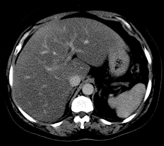
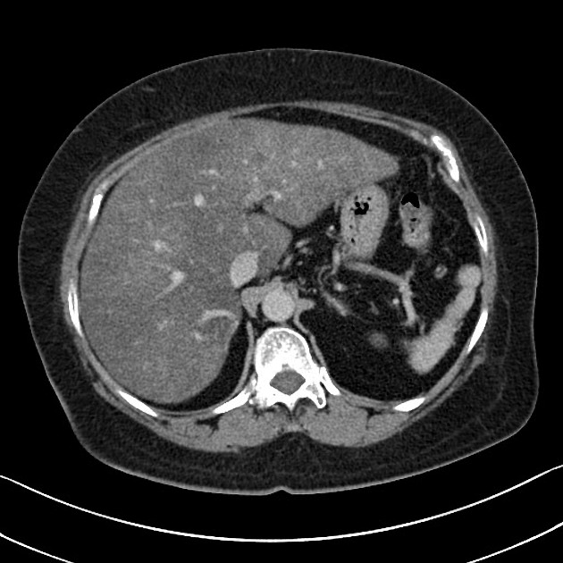
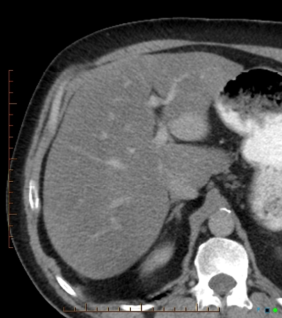
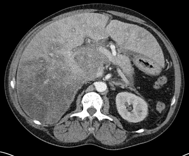
Unusual patterns such as multifocal nodular hepatic steatosis can be mistaken for metastases 9.
Ultrasound
Ultrasound B-mode imaging is preferred as the first-line diagnostic modality for hepatic steatosis because of its wide availability, low cost, non-invasive, does not expose to ionizing radiation, repeatable, and well accepted by patients 2.
The degree of diffuse hepatic steatosis can be graded according to several different scoring systems, including the Hamaguchi score, US-FLI score, and hepatorenal sonographic index 2.
Attenuation imaging is also another new method which can be used for quantifying degree of steatosis.
CT
Liver attenuation of < 40-45 HU on non-contrast CT identifies moderate hepatic steatosis (> 20-33% at biopsy) with a sensitivity of 82% and specificity of 94% 7. Of course other factors affect attenuation, such as edema, protein content and iron or iodine deposition.
MRI
Proton density fat fraction is accurate and correlates well with biopsy 8.


 Unable to process the form. Check for errors and try again.