
Hydronephrosis
Citation, DOI, disclosures and article data
At the time the article was created Bruno Di Muzio had no recorded disclosures.
View Bruno Di Muzio's current disclosuresAt the time the article was last revised Tariq Walizai had no financial relationships to ineligible companies to disclose.
View Tariq Walizai's current disclosures- Hydronephroses
- Hydroureteronephroses
- Ureterohydronephroses
- Ureterohydronephrosis
- Hydroureteronephrosis
- Calyceal dilatation
- Calicectasis
- Caliectasis
- Pelvicalyceal dilatation
- Pelvocaliectasis
- Pyelocaliectasis
- Pelvic dilatation
- Pelviectasis
- Pyelectasis
- Ureteropelvicalyceal dilatation
- Ureteral and pelvicalyceal dilatations
- Ureteral dilatation
- Ureterectasis
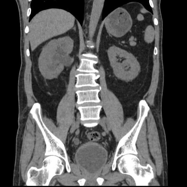
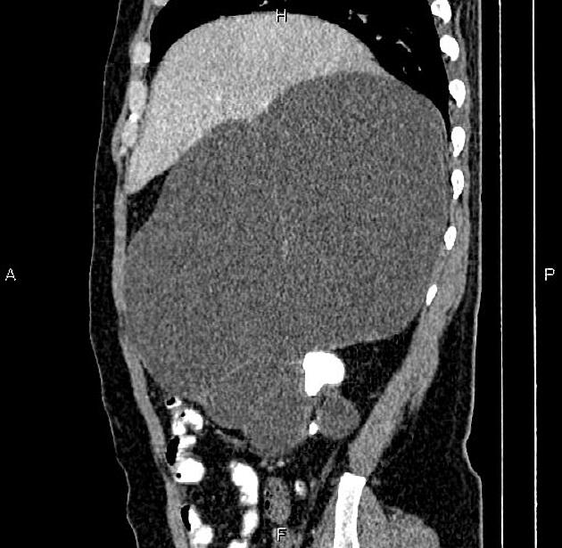
Hydronephrosis (plural: hydronephroses) is defined as dilatation of the urinary collecting system of the kidney (the calyces, the infundibula, and the pelvis) 1.
Hydronephrosis in fetuses and newborns has specific causes that are covered in a separate article.
On this page:
Terminology
The term hydroureteronephrosis (or hydronephroureterosis) may be used when the dilatation occurs in the presence of hydroureter.
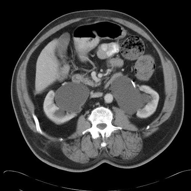
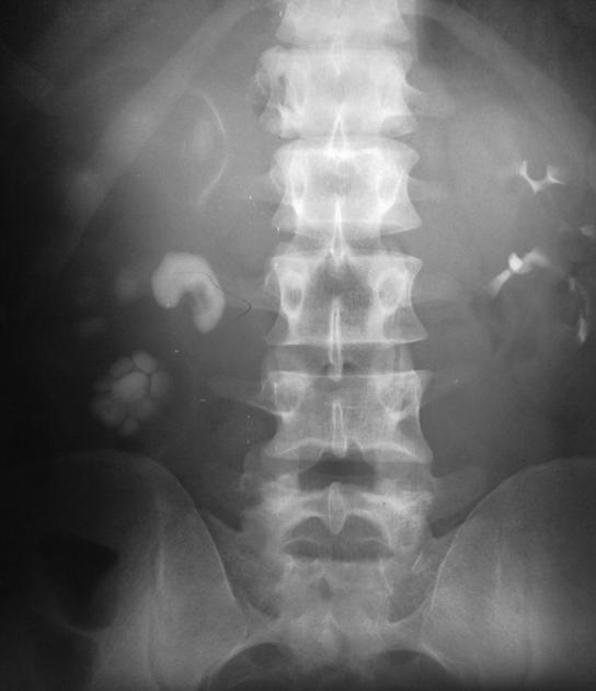
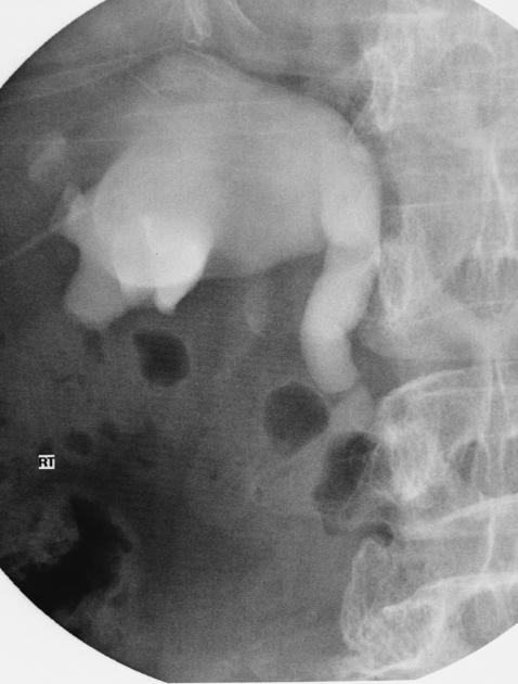
Radiographic features
Following the identification of hydronephrosis, appropriate further investigations must be undertaken to establish an underlying cause, with potential etiologies including everything from urolithiasis, pelviureteric junction obstruction, malignancy such as cervical cancer, and retroperitoneal fibrosis.
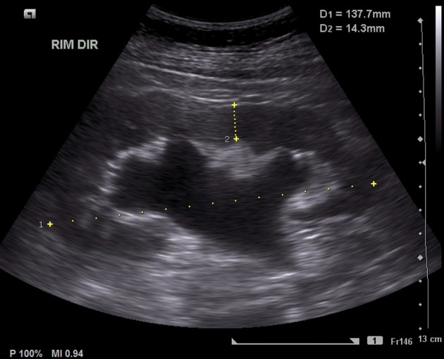
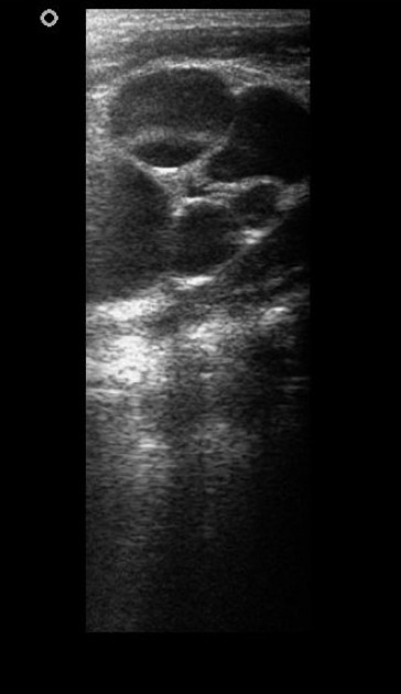
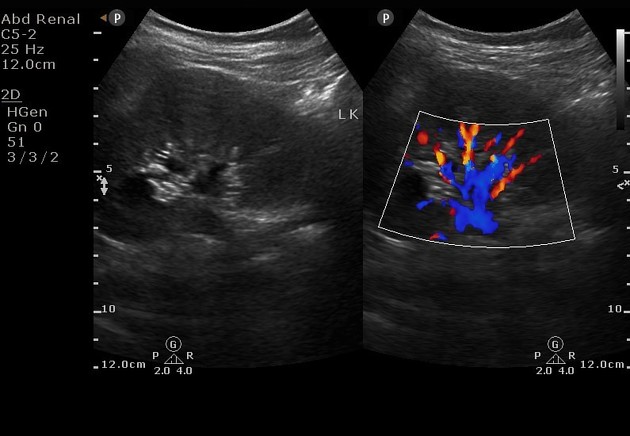
Ultrasound
Ultrasound imaging of hydronephrosis will demonstrate a dilated pelvicalcyceal system. The severity is often classified into mild, moderate or severe hydronephrosis. Thinning of the renal cortex in the context of hydronephrosis usually implies chronicity. Of note, bladder outflow obstruction (or simply a very full bladder) may result in a bilaterally prominent pelvicalyceal system. This can be assessed by rescanning the kidneys post-void to assess for change in the degree of pelvicalyceal dilatation.
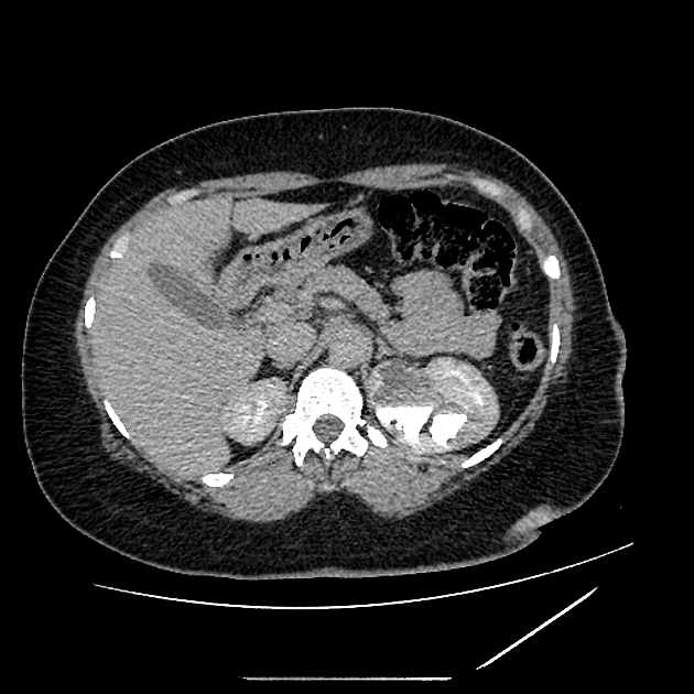
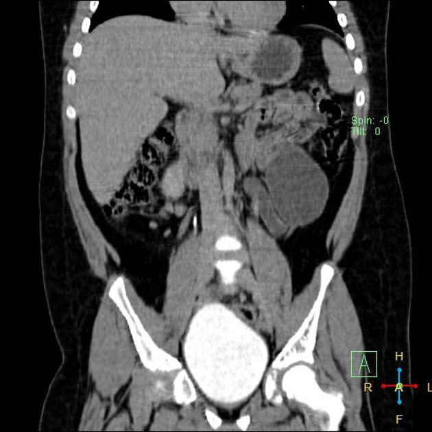
CT
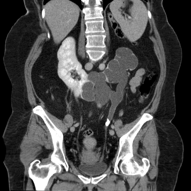
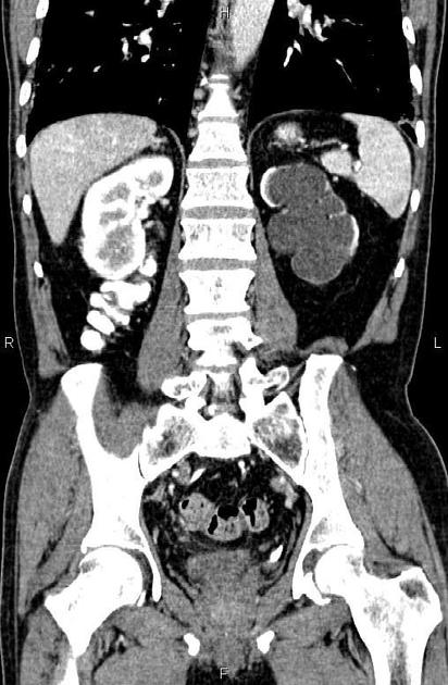
CT will readily show hydronephrosis, and can also help identify the cause.
Unenhanced CT is often used to look for urinary tract calculi.
Contrast enhanced CT in the portal venous phase can help to delineate other causes of hydronephrosis, such as retroperitoneal fibrosis and pelvic malignancies.
Delayed phase contrast enhanced CT imaging is useful for intrinsic assessment of the collecting system, and can more clearly demonstrate ureteric strictures or carcinomas, bladder malignancies and non-calcified stones.
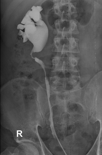
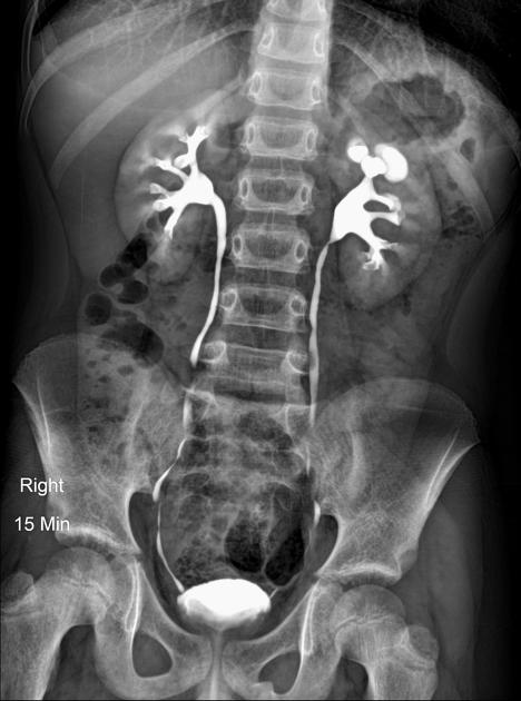
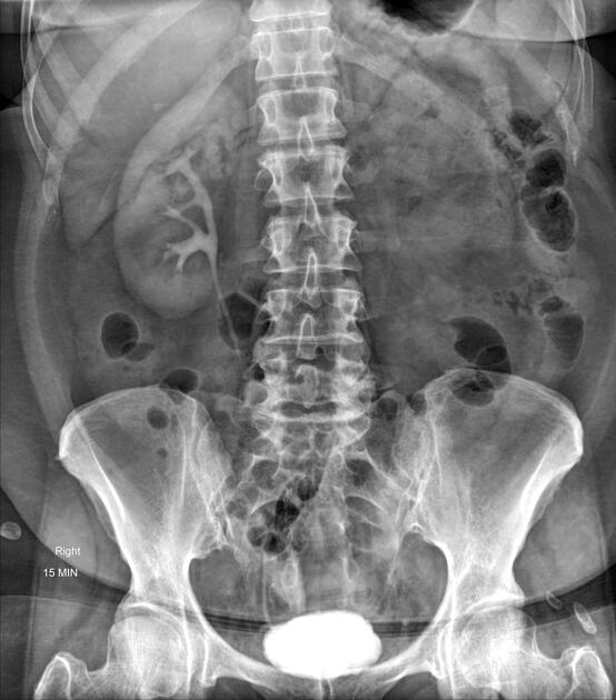
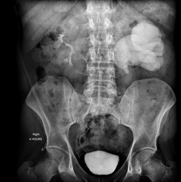
Nuclear medicine
A nuclear medicine diuretic renogram may be performed to assess for obstruction of urine and differentiate from other causes such as an extra-renal pelvis or parapelvic cysts.
The radiologist may also play a part in procedures to treat the harmful effects of uncorrected hydronephrosis on renal function, such as placement of a percutaneous nephrostomy tube or antegrade ureteric stent insertion.
ADVERTISEMENT: Supporters see fewer/no ads
Differential diagnosis
See also
References
- 1. Robins S & Fischmann J. Hydronephrosis; a Radiologic Classification Based on Anatomical Variations. Radiology. 1948;50(5):632-8. doi:10.1148/50.5.632 - Pubmed
Incoming Links
- Voiding cystourethrography
- Urolithiasis
- Transitional cell carcinoma (urinary tract)
- Kidney ultrasound
- Prostate cancer
- Renal lymphoma
- Inflammatory abdominal aortic aneurysm
- Calyceal crescent sign (intravenous pyelogram)
- Renal tuberculosis
- Fetal conditions associated with maternal diabetes
- Haematometrocolpos
- CT urography (protocol)
- SFU grading system of hydronephrosis
- Frozen pelvis
- Radiation and chemotherapy induced cystitis
- Xanthogranulomatous pyelonephritis
- Medical abbreviations and acronyms (H)
- Hydroureter
- Transitional cell carcinoma (renal pelvis)
- Renal transplant ultrasound
- Retroperitoneal fibrosis
- Ketamine bladder
- Ureteropelvic junction obstruction
- Urinary bladder tumour - pulmonary pseudometastasis
- Ureterocele
- Pelviureteric junction obstruction
- Incidental bilateral vesicoureteral reflux
- Bladder outlet obstruction with bilateral pyelonephritis
- Ureteral stricture
- Obstructed primary megaureter
- Peripelvic cysts and pseudohydronephrosis
- Migrated double J stent
- Renal artery aneurysm causing focal hydronephrosis
- Staghorn stone
- Pelviureteric junction obstruction
- Obstructed hydrocalyx
- Vesicoureteral reflux (cystourethrography)
- Ureteric calculus and hydronephrosis (x-ray)
- Crossed fused renal ectopia
- Bilateral ureteral compression by iliac arteries
Related articles: Pathology: Genitourinary
- obstetrics
-
first trimester
- ultrasound findings in early pregnancy
- embryo/fetus
- beta-hCG levels
- confirming intrauterine gestation
- pregnancy of unknown location (PUL)
- first trimester vaginal bleeding
- early structural scan
- aneuploidy testing
-
second trimester
- fetal biometry
- amniotic fluid volume
- fetal morphology assessment
- soft markers
- amnioreduction
- Doppler ultrasound
- nuchal translucency
- 11-13 weeks antenatal scan
- chorionic villus sampling (CVS) and amniocentesis
- other
- placenta
- placental anatomy
- placental developmental abnormalities
- placenta previa
- spectrum of abnormal placental villous adherence
- abnormalities of cord insertion
- abruptio placentae
- placental pathology
- vascular pathologies of placenta
- placental infections
- placental masses
- molar pregnancy
- twin placenta
- miscellaneous
-
first trimester
- gynecology
- acute pelvic pain
- chronic pelvic pain
- uterus
- ovaries
- ovarian follicle
- ovarian torsion
- pelvic inflammatory disease
- ovarian cysts and masses
- paraovarian cyst
- polycystic ovaries
- ovarian hyperstimulation syndrome
- post-hysterectomy ovary
- cervix
- fallopian tube
- other
- male genital tract
- prostate gland
- transrectal ultrasound
- prostate tumors
- infections of the prostate
-
prostatitis
- acute bacterial prostatitis
-
chronic prostatitis
- chronic bacterial prostatitis
- chronic prostatitis and chronic pelvic pain syndrome (CPPS)
- asymptomatic inflammatory prostatitis
- granulomatous prostatitis
- emphysematous prostatitis
- prostatic abscess
-
prostatitis
- benign prostatic hypertrophy
- cystic lesions of the prostate
- prostatic calcification
- prostatic infarction
- testes
-
unilateral testicular lesion
- testicular torsion
- orchitis
- testicular trauma
-
germ cell tumors of the testis
- testicular seminoma
-
non seminomatous germ cell tumors
- mixed germ cell tumor
- yolk sac tumor (endodermal sinus tumor)
- embryonal cell carcinoma
- choriocarcinoma
- testicular teratoma
- testicular epidermoid (teratoma with ectodermal elements only)
- burned out testis tumor
- sex cord / stromal tumors of the testis
- testicular cyst
- testicular lymphoma
- bilateral testicular lesion
- paratesticular lesions
- epididymis
- other
- polyorchidism
- cryptorchidism
- tubular ectasia of the rete testis
- cystadenoma of the rete testis
- testicular sarcoidosis
- testicular tuberculosis
- spermatic cord
- fibrous pseudotumor of the scrotum
- scrotal leiomyosarcoma
- testicular adrenal rest tumors (TARTs)
- tunica vaginalis testis mesothelioma
- splenogonadal fusion
- testicular vasculitis
- abnormal testicular Doppler flow (differential)
-
unilateral testicular lesion
- penis
- prostate gland
- KUB
- kidneys
- normal renal anatomy
- hydronephrosis
- urolithiasis
- renal masses
- renal cystic disease
- renal infection
- vascular
- trauma
- ureter
- normal ureter anatomy
- ureteral stricture
- ureteral dilatation
- ureteral anomalies
- ureteral tumors
- ureteral trauma
- other
- bladder
- kidneys


 Unable to process the form. Check for errors and try again.