
Inferior nasal concha
Updates to Article Attributes
The inferior nasal concha or turbinate is one of the conchae in the nose.
Gross anatomy
It extends horizontally along the lateral wall of the nasal cavity and consists of a lamina of spongy bone, curled upon itself like a scroll. Each inferior nasal concha is considered a facial pair of facial bones since they arise from the maxillae bones and projects horizontally into the nasal cavity.
Superior to inferior nasal concha are the middle nasal concha and superior nasal concha which arise from the cranial portion of the skull. Hence, these two are considered as a part of the cranial bones.
Surfaces
The medial surface is convex, perforated by numerous apertures, and traversed by longitudinal grooves for the lodgement of vessels. The lateral surface is concave, and forms part of the inferior meatus.
Borders
Its upper border is thin, irregular, and connected to various bones along the lateral wall of the nasal cavity. The inferior border is free, thick, and cellular in structure, more especially in the middle of the bone.
The upper border may be divided into three portions:
- the anterior articulates with the conchal crest of the maxilla
- the posterior with the conchal crest of the palatine
- the middle portion presents three well-marked processes, which vary much in their size and form
Ossification
Development
The inferior nasal concha is ossified from a single centercentre, which appears about the fifth month of fetal life in the lateral wall of the cartilaginous nasal capsule.
-<p>The <strong>inferior nasal concha</strong> is one of the <a href="/articles/nasal-concha">conchae</a> in the nose. It extends horizontally along the lateral wall of the nasal cavity and consists of a lamina of spongy bone, curled upon itself like a scroll. Each inferior nasal concha is considered a facial pair of bones since they arise from the <a href="/articles/maxilla">maxillae bones</a> and projects horizontally into the nasal cavity.</p><p>Superior to inferior nasal concha are the <a href="/articles/middle-nasal-concha">middle nasal concha</a> and <a href="/articles/superior-nasal-concha">superior nasal concha</a> which arise from the cranial portion of the skull. Hence, these two are considered as a part of the cranial bones.</p><h3>Surfaces</h3><p>The medial surface is convex, perforated by numerous apertures, and traversed by longitudinal grooves for the lodgement of vessels. The lateral surface is concave, and forms part of the inferior meatus.</p><h3>Borders</h3><p>Its upper border is thin, irregular, and connected to various bones along the lateral wall of the nasal cavity. The inferior border is free, thick, and cellular in structure, more especially in the middle of the bone.</p><p>The upper border may be divided into three portions:</p><ul>- +<p>The <strong>inferior nasal concha</strong> or <strong>turbinate</strong> is one of the <a href="/articles/nasal-concha">conchae</a> in the <a title="Nose" href="/articles/nose">nose</a>.</p><h4>Gross anatomy</h4><p>It extends horizontally along the lateral wall of the <a title="Nasal cavity" href="/articles/nasal-cavity">nasal cavity</a> and consists of a lamina of spongy bone, curled upon itself like a scroll. Each inferior nasal concha is considered a pair of <a title="Facial bones" href="/articles/facial-bones">facial bones</a> since they arise from the <a href="/articles/maxilla">maxillae bones</a> and projects horizontally into the nasal cavity.</p><p>Superior to inferior nasal concha are the <a href="/articles/middle-nasal-concha">middle nasal concha</a> and <a href="/articles/superior-nasal-concha">superior nasal concha</a> which arise from the cranial portion of the skull. Hence, these two are considered as a part of the cranial bones.</p><h5>Surfaces</h5><p>The medial surface is convex, perforated by numerous apertures, and traversed by longitudinal grooves for the lodgement of vessels. The lateral surface is concave and forms part of the inferior meatus.</p><h5>Borders</h5><p>Its upper border is thin, irregular, and connected to various bones along the lateral wall of the nasal cavity. The inferior border is free, thick, and cellular in structure, more especially in the middle of the bone.</p><p>The upper border may be divided into three portions:</p><ul>
-</ul><h3>Ossification</h3><p>The inferior nasal concha is ossified from a single center, which appears about the fifth month of fetal life in the lateral wall of the cartilaginous nasal capsule.</p>- +</ul><h4>Development</h4><p>The inferior nasal concha is ossified from a single centre, which appears about the fifth month of fetal life in the lateral wall of the cartilaginous nasal capsule.</p>
References changed:
- 1. Henry Gray, Susan Standring, B. K. B. Berkovitz. Gray's Anatomy. (2005) ISBN: 9780443071690 - <a href="http://books.google.com/books?vid=ISBN9780443071690">Google Books</a>
- <a href="http://en.wikipedia.org/wiki/Inferior_nasal_conchae">Wikipedia: inferior nasal concha</a>
- Gray's Anatomy 39th Edition, Elsevier
Tags changed:
- refs
Image 3 Diagram ( update )
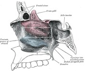
Image 5 Diagram ( update )


 Unable to process the form. Check for errors and try again.