
Lisfranc injury
Updates to Article Attributes
Lisfranc injuries,also called Lisfranc fracture-dislocations, are the most common type of dislocation involving the foot and correspond to the dislocation of the articulation of the tarsus with the metatarsal bases.
Pathology
Anatomy
The Lisfranc joint articulates the tarsus with the metatarsal bases, whereby the first three metatarsals articulate respectively with the three cuneiforms, and the 4th and 5th metatarsals with the cuboid.
The Lisfranc ligament is a strong band attaching the medial cuneiform to the 2nd metatarsal base on the foot's plantar aspect. Its integrity is crucial to the stability of the Lisfranc joint.
Aetiology
Injury mechanisms are varied and include direct crush injury or an indirect load onto a plantarflexed foot 3. Tarsometatarsal dislocation may also occur in the diabetic neuropathic joint (Charcot).
Subtypes
There are several types of Lisfranc fracture-dislocation:
- homolateral: a homolateral injury is a lateral displacement of the 1st to 5th metatarsals or of 2nd to 5th metatarsals where the 1st MTP joint remains congruent
- divergent: a divergent injury is a lateral dislocation of the 2nd to 5th metatarsals with medial dislocation of the 1st metatarsal
- isolated: this involves one or two metatarsals that dislocate dorsally in isolation
Radiographic features
Plain radiograph/CT
These injuries are well demonstrated on the standard views of the foot. Still, subtle injuries may be missed and require further imaging such as CT, MRI or radiographic stress views with forefoot abduction. CT is, however, favoured as it will also demonstrate unsuspected associated fractures.
The key finding is malalignment of the second tarsometatarsal joint, such as lateral displacement of the second metatarsal base on AP view and/or dorsal step-off sign on lateral view 10. An additional abnormality is diastasis >2 mm between the first1st and second2nd metatarsal bases 10.
Other possible findings are malalignment between the lateral border of the base of the 1st metatarsal and the lateral border of the medial cuneiform; malalignment between the medial border of the base of the 4th metatarsal and the cuboid (on the oblique view); increased distance between the medial cuneiform and the 2nd metatarsal; and increased distance between the medial and intermediate cuneiforms (C2) 13.
Associated fractures most often occur at the base of the second metatarsal, seen as the fleck sign. They may also be seen in the 3rd metatarsal, 1st or 2nd cuneiform, or navicular bones.
If the diagnosis is in doubt, it may be useful to obtain weight-bearing x-rays and comparison views of the contralateral side 11.
Ultrasound
Useful for assessing the ligamentous injury. Non-visualisation of the dorsal C1-M2 ligament and a C1-M2 distance >2.5 mm is indirectly indicative of a Lisfranc ligament tear 5.
Dynamic evaluation with weight-bearing may show widening of the space between C1 and M2.
MRI
Again may be useful for assessing ligamentous injury, especially when there is a high clinical concern with routine radiographs being inconclusive 7.
Treatment and prognosis
Treatment may be non-operative or operative, with the aim being to have a painless, plantigrade and stable foot 12.
Indications for non-operative treatment include undisplaced injuries that are stable with weight-bearing or poor surgical candidates such as non-ambulatory patients, patients with significant comorbidities that have high risk for complications (e.g. severe vascular disease, peripheral neuropathy) or pre-existing inflammatory arthritis 12.
The indication for operative management is an unstable injury. These can be divided into joint saving or joint sacrificing. Joint saving surgery includes temporary fixation whilst awaiting definitive management and ORIF. Joint sacrificing surgery is either arthrodesis of the 1st, 2nd and 3rd tarsometatarsal joints or midfoot arthrodesis 12.
Internal fixation is the most common treatment.
Complications
The most common complications of ankle and foot fractures are non-union and post-traumatic arthritis. Although conventional radiography can usually demonstrate these complications' features, CT is the better technique for delineating their details.
History and etymology
It is named after Jacques Lisfranc De Saint Martin (1790-1847), the chief of surgery at the Hôpital de la Pitie in Paris 2.
-</ul><h4>Radiographic features</h4><h5>Plain radiograph/CT</h5><p>These injuries are well demonstrated on the standard views of the foot. Still, subtle injuries may be missed and require further imaging such as CT, MRI or radiographic stress views with forefoot abduction. CT is, however, favoured as it will also demonstrate unsuspected associated fractures.</p><p>The key finding is malalignment of the second tarsometatarsal joint, such as lateral displacement of the second metatarsal base on AP view and/or dorsal <a href="/articles/step-off-sign">step-off sign</a> on lateral view <sup>10</sup>. An additional abnormality is diastasis >2 mm between the first and second metatarsal bases <sup>10</sup>.</p><p>Associated fractures most often occur at the base of the second metatarsal, seen as the <a href="/articles/fleck-sign-foot">fleck sign</a>. They may also be seen in the 3<sup>rd</sup> metatarsal, 1<sup>st </sup>or 2<sup>nd</sup> cuneiform, or navicular bones. </p><p>If the diagnosis is in doubt, it may be useful to obtain weight-bearing x-rays and comparison views of the contralateral side <sup>11</sup>.</p><h5>Ultrasound</h5><p>Useful for assessing the ligamentous injury. Non-visualisation of the dorsal C1-M2 ligament and a C1-M2 distance >2.5 mm is indirectly indicative of a Lisfranc ligament tear <sup>5</sup>.</p><p>Dynamic evaluation with weight-bearing may show widening of the space between C1 and M2.</p><h5>MRI</h5><p>Again may be useful for assessing ligamentous injury, especially when there is a high clinical concern with routine radiographs being inconclusive <sup>7</sup>. </p><h4>Treatment and prognosis</h4><p>Treatment may be non-operative or operative, with the aim being to have a painless, plantigrade and stable foot <sup>12</sup>.</p><p>Indications for non-operative treatment include undisplaced injuries that are stable with weight-bearing or poor surgical candidates such as non-ambulatory patients, patients with significant comorbidities that have high risk for complications (e.g. severe vascular disease, peripheral neuropathy) or pre-existing inflammatory arthritis <sup>12</sup>.</p><p>The indication for operative management is an unstable injury. These can be divided into joint saving or joint sacrificing. Joint saving surgery includes temporary fixation whilst awaiting definitive management and ORIF. Joint sacrificing surgery is either arthrodesis of the 1st, 2nd and 3rd tarsometatarsal joints or midfoot arthrodesis <sup>12</sup>.</p><p>Internal fixation is the most common treatment. </p><h5>Complications</h5><p>The most common complications of ankle and foot fractures are non-union and post-traumatic arthritis. Although conventional radiography can usually demonstrate these complications' features, CT is the better technique for delineating their details.</p><h4>History and etymology</h4><p>It is named after <strong>Jacques Lisfranc De Saint Martin</strong> (1790-1847), the chief of surgery at the Hôpital de la Pitie in Paris <sup>2</sup>.</p>- +</ul><h4>Radiographic features</h4><h5>Plain radiograph/CT</h5><p>These injuries are well demonstrated on the standard views of the foot. Still, subtle injuries may be missed and require further imaging such as CT, MRI or radiographic stress views with forefoot abduction. CT is, however, favoured as it will also demonstrate unsuspected associated fractures.</p><p>The key finding is malalignment of the second tarsometatarsal joint, such as lateral displacement of the second metatarsal base on AP view and/or dorsal <a href="/articles/step-off-sign">step-off sign</a> on lateral view <sup>10</sup>. An additional abnormality is diastasis >2 mm between the 1<sup>st</sup> and 2<sup>nd</sup> metatarsal bases <sup>10</sup>.</p><p>Other possible findings are malalignment between the lateral border of the base of the 1<sup>st</sup> metatarsal and the lateral border of the medial cuneiform; malalignment between the medial border of the base of the 4<sup>th</sup> metatarsal and the cuboid (on the oblique view); increased distance between the medial cuneiform and the 2<sup>nd</sup> metatarsal; and increased distance between the medial and intermediate cuneiforms (C2) <sup>13</sup>.</p><p>Associated fractures most often occur at the base of the second metatarsal, seen as the <a href="/articles/fleck-sign-foot">fleck sign</a>. They may also be seen in the 3<sup>rd</sup> metatarsal, 1<sup>st </sup>or 2<sup>nd</sup> cuneiform, or navicular bones. </p><p>If the diagnosis is in doubt, it may be useful to obtain weight-bearing x-rays and comparison views of the contralateral side <sup>11</sup>.</p><h5>Ultrasound</h5><p>Useful for assessing the ligamentous injury. Non-visualisation of the dorsal C1-M2 ligament and a C1-M2 distance >2.5 mm is indirectly indicative of a Lisfranc ligament tear <sup>5</sup>.</p><p>Dynamic evaluation with weight-bearing may show widening of the space between C1 and M2.</p><h5>MRI</h5><p>Again may be useful for assessing ligamentous injury, especially when there is a high clinical concern with routine radiographs being inconclusive <sup>7</sup>. </p><h4>Treatment and prognosis</h4><p>Treatment may be non-operative or operative, with the aim being to have a painless, plantigrade and stable foot <sup>12</sup>.</p><p>Indications for non-operative treatment include undisplaced injuries that are stable with weight-bearing or poor surgical candidates such as non-ambulatory patients, patients with significant comorbidities that have high risk for complications (e.g. severe vascular disease, peripheral neuropathy) or pre-existing inflammatory arthritis <sup>12</sup>.</p><p>The indication for operative management is an unstable injury. These can be divided into joint saving or joint sacrificing. Joint saving surgery includes temporary fixation whilst awaiting definitive management and ORIF. Joint sacrificing surgery is either arthrodesis of the 1st, 2nd and 3rd tarsometatarsal joints or midfoot arthrodesis <sup>12</sup>.</p><p>Internal fixation is the most common treatment. </p><h5>Complications</h5><p>The most common complications of ankle and foot fractures are non-union and post-traumatic arthritis. Although conventional radiography can usually demonstrate these complications' features, CT is the better technique for delineating their details.</p><h4>History and etymology</h4><p>It is named after <strong>Jacques Lisfranc De Saint Martin</strong> (1790-1847), the chief of surgery at the Hôpital de la Pitie in Paris <sup>2</sup>.</p>
References changed:
- 13. Sripanich Y, Weinberg M, Krähenbühl N et al. Imaging in Lisfranc Injury: A Systematic Literature Review. Skeletal Radiol. 2020;49(1):31-53. <a href="https://doi.org/10.1007/s00256-019-03282-1">doi:10.1007/s00256-019-03282-1</a> - <a href="https://www.ncbi.nlm.nih.gov/pubmed/31368007">Pubmed</a>
Image ( update )
Image ( update )
Image ( update )
Image ( update )
Image ( update )
Image 5 Diagram ( create )

Image 7 Diagram ( update )

Image 8 Diagram ( update )

Image 9 X-ray (Frontal) ( update )

Image 10 X-ray (Oblique) ( update )

Image 11 X-ray (Frontal) ( update )

Image 12 Annotated image (Frontal) ( update )

Image 13 X-ray (Frontal) ( update )

Image 14 X-ray ( update )
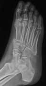
Image 15 X-ray (Frontal) ( update )

Image 16 X-ray ( update )

Image 17 X-ray (Frontal) ( update )

Image 18 X-ray (Oblique) ( update )
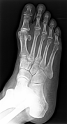
Image 19 CT (bone window) ( update )


 Unable to process the form. Check for errors and try again.