
Musculoskeletal imaging (dual-energy CT)
Updates to Article Attributes
Dual-energy CT has a number of clinical applications in the assessment of the musculoskeletal system particularly in the realm of artefact reduction and material composition, .
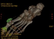
Detection of bone marrow oedema
Similar to the concept of using virtual non-contrast imaging, virtual non-calcium images can be created using a three-material decomposition technique, to subtract the high attenuating trabecular bone and allow for assessment of bone marrow oedema 11. Using histhis technique one can appreciate areas of increased areas of attenuation within the bone marrow. Using this post-processing technique one and hence can better appreciate occult fractures that lack or possess a limited cortical component. Although it should be noted that bone marrow oedema is not pathognomonic for a subtle fracture.
Metal artefact reduction
Conventional CT suffers from decreased image quality when faced with metal implants, metal in the CT scan will lead to artefacts such as beam gardeninghardening, excessive beam attenuation, photon starvation and overall degradation of the image. A method to overcome this is a virtual monochromatic energy spectrum reconstruction from a dual-energy acquisition, whereby a higher energy data set can be created (often around 105 and 133 kV 2) to decrease attenuation (increase penetration [virtually]) and overall decrease metal related artefact 2.
Gout
Traditionally gout has been diagnosed via plain radiography and joint aspiration. Dual-energy CT has become a reliable, non-invasive technique to make this diagnosis 2,3. Taking into account the differing attenuation values of material, it is possible to detect and quantify urate deposits within CT scans. Furthermore, it is a useful rule post-processing technique to determine if a region of interest is, infact, tophaceous gout or an infection of mass/mass.
-<p>Dual-energy CT has a number of clinical applications in the assessment of the <strong>musculoskeletal system </strong>particularly in the realm of artefact reduction and material composition, </p><h4>Detection of bone marrow oedema </h4><p>Similar to the concept of using<a title="Virtual non-contrast imaging" href="/articles/virtual-non-contrast-imaging"> virtual non-contrast imaging</a>, virtual non-calcium images can be created using a three-material decomposition technique, to subtract the high attenuating trabecular bone and allow for assessment of <a title="Bone marrow oedema" href="/articles/bone-marrow-oedema">bone marrow oedema</a> 1. Using his technique one can appreciate areas of increased areas of attenuation within the bone marrow. Using this post-processing technique one can better appreciate occult fractures that lack or possess a limited cortical component. Although it should be noted that bone marrow oedema is not pathognomonic for a subtle fracture. </p><h4>Metal artefact reduction</h4><p>Conventional CT suffers from decreased image quality when faced with metal implants, metal in the CT scan will lead to artefacts such as beam gardening excessive beam attenuation, photon starvation and overall degradation of the image. A method to overcome this is a virtual monochromatic energy spectrum reconstruction from a dual-energy acquisition, whereby a higher energy data set can be created (often around 105 and 133 kV <sup>2</sup>) to decrease attenuation and overall decrease metal related artefact <sup>2</sup>. </p><h4>Gout </h4><p>Traditionally gout has been diagnosed via plain radiography and joint aspiration. Dual-energy CT has become a reliable, non-invasive technique to make this diagnosis <sup>2,3</sup>. Taking into account the differing attenuation values of material, it is possible to detect and quantify urate deposits within CT scans. Furthermore, it is a useful rule post-processing technique to determine if a region of interest is, infact, tophaceous gout or an infection of mass. </p>- +<p>Dual-energy CT has a number of clinical applications in the assessment of the <strong>musculoskeletal system </strong>particularly in the realm of artefact reduction and material composition.</p><h4>Detection of bone marrow oedema</h4><p>Similar to the concept of using<a href="/articles/virtual-non-contrast-imaging"> virtual non-contrast imaging</a>, virtual non-calcium images can be created using a three-material decomposition technique, to subtract the high attenuating trabecular bone and allow for assessment of <a href="/articles/bone-marrow-oedema">bone marrow oedema</a> <sup>1</sup>. Using this technique one can appreciate areas of increased attenuation within the bone marrow and hence can better appreciate occult fractures that lack or possess a limited cortical component. Although it should be noted that bone marrow oedema is not pathognomonic for a subtle fracture. </p><h4>Metal artefact reduction</h4><p>Conventional CT suffers from decreased image quality when faced with metal implants, metal in the CT scan will lead to artefacts such as <a href="/articles/beam-hardening">beam hardening</a>, excessive beam attenuation, <a href="/articles/photon-starvation-2">photon starvation</a> and overall degradation of the image. A method to overcome this is a virtual monochromatic energy spectrum reconstruction from a dual-energy acquisition, whereby a higher energy data set can be created (often around 105 and 133 kV <sup>2</sup>) to decrease attenuation (increase penetration [virtually]) and overall decrease metal related artefact <sup>2</sup>. </p><h4>Gout </h4><p>Traditionally gout has been diagnosed via plain radiography and joint aspiration. Dual-energy CT has become a reliable, non-invasive technique to make this diagnosis <sup>2,3</sup>. Taking into account the differing attenuation values of material, it is possible to detect and quantify urate deposits within CT scans. Furthermore, it is a useful rule post-processing technique to determine if a region of interest is, infact, tophaceous gout or an infection/mass. </p>
References changed:
- 1. Babina Gosangi, Jacob C. Mandell, Michael J. Weaver, Jennifer W. Uyeda, Stacy E. Smith, Aaron D. Sodickson, Bharti Khurana. Bone Marrow Edema at Dual-Energy CT: A Game Changer in the Emergency Department. (2020) RadioGraphics. 40 (3): 859-874. <a href="https://doi.org/10.1148/rg.2020190173">doi:10.1148/rg.2020190173</a> - <a href="https://www.ncbi.nlm.nih.gov/pubmed/32364883">Pubmed</a> <span class="ref_v4"></span>
- 2. Paul I. Mallinson, Tyler M. Coupal, Patrick D. McLaughlin, Savvas Nicolaou, Peter L. Munk, Hugue A. Ouellette. Dual-Energy CT for the Musculoskeletal System. (2016) Radiology. 281 (3): 690-707. <a href="https://doi.org/10.1148/radiol.2016151109">doi:10.1148/radiol.2016151109</a> - <a href="https://www.ncbi.nlm.nih.gov/pubmed/27870622">Pubmed</a> <span class="ref_v4"></span>
- 3. Hong Chou, Teck Yew Chin, Wilfred C. G. Peh. Dual‐energy CT in gout – A review of current concepts and applications. (2017) Journal of Medical Radiation Sciences. 64 (1): 41. <a href="https://doi.org/10.1002/jmrs.223">doi:10.1002/jmrs.223</a> - <a href="https://www.ncbi.nlm.nih.gov/pubmed/28238226">Pubmed</a> <span class="ref_v4"></span>
Tags changed:
- rg_40_3_edit
Sections changed:
- Radiography
Systems changed:
- Musculoskeletal
Image 1 CT (Edema map) ( create )

Image 2 CT ( create )


 Unable to process the form. Check for errors and try again.