
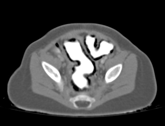
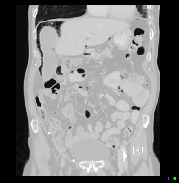
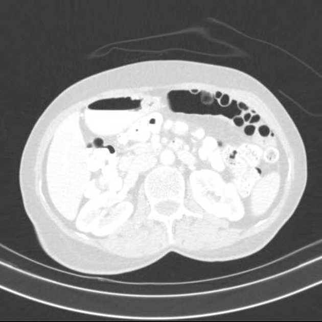
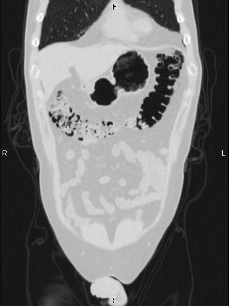
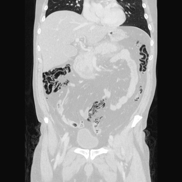
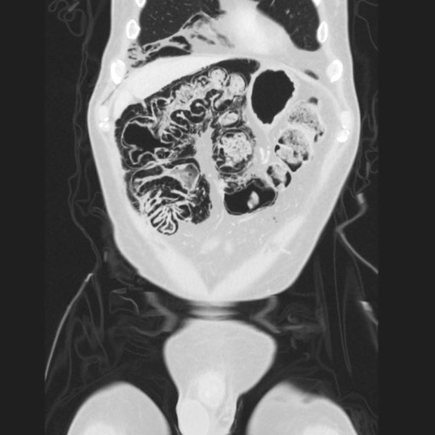
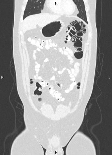
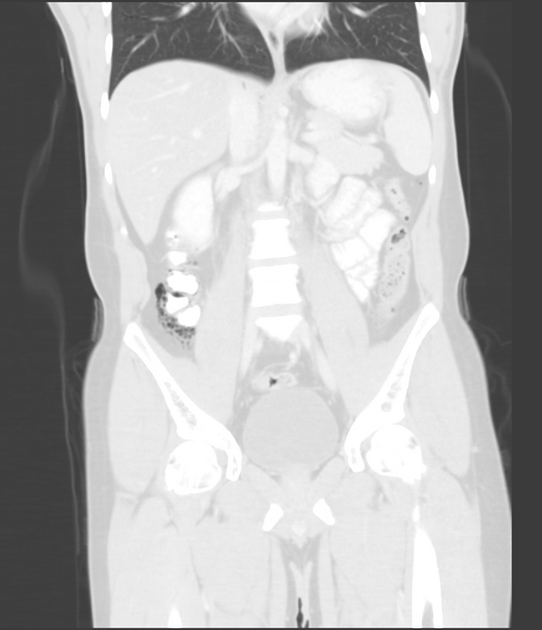
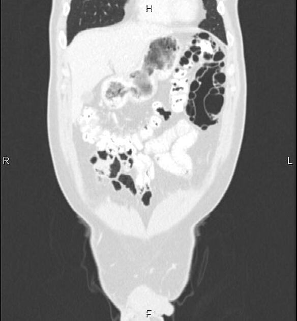
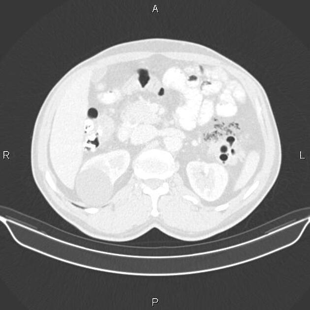
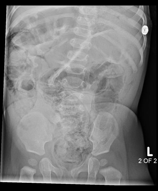
Pneumatosis coli is a descriptive sign presenting radiographically as intramural gas limited to the colonic wall.
On this page:
Terminology
There are different terminologies in the medical literature, such as pneumatosis intestinalis, pneumatosis coli, and pneumatosis cystoides intestinalis. Pneumatosis intestinalis is used when only the small bowel wall is involved. Pneumatosis cystoides intestinalis (or coli) is descriptive for multiple gaseous cysts along the bowel wall.
There are 2 types of pneumatosis coli based on morphology 7:
pneumatosis cystoides coli: usually due to benign entities
pneumatosis linearis coli: secondary to ischemic and necrotic bowel
Pathology
In the pediatric population, it is most frequently seen in premature infants 1. Although symptoms are relatively mild, they are the same as those seen in early (stage I) necrotizing enterocolitis.
In adults, it can have both benign and life-threatening causes.
Benign pneumatosis can be caused by a variety of reasons such as pulmonary disease, systemic disease (scleroderma, lupus, AIDS), intestinal inflammation, iatrogenic/procedures, medications, and organ transplantation 5.
Life-threatening pneumatosis can be caused by intestinal ischemia, obstruction, enteritis/colitis, toxic caustic ingestion, toxic megacolon, organ transplantation, and collagen vascular disease 5.
Radiographic features
The following are imaging features of clinically worrisome pneumatosis 6:
soft tissue bowel wall thickening
free intraperitoneal fluid
lesser extent of pneumatosis (more extensive pneumatosis is more commonly benign)
peri-intestinal soft-tissue stranding
abnormal bowel wall enhancement
atherosclerosis and vascular occlusion
Pneumoperitoneum and pneumoretroperitoneum can be seen with both idiopathic and ischemic pneumatosis 6.
Differential diagnosis
Pseudopneumatosis (mimics) include:
gas trapped between bowel wall and luminal contents
gas trapped by opposing mucosal folds
gas bubbles adherent to the bowel wall


 Unable to process the form. Check for errors and try again.