
Pneumatosis cystoides intestinalis
Updates to Article Attributes
Pneumatosis cystoides intestinalis (PCI) is a radiographic/descriptive entity characterized by the presence of multiple gas filled cysts in the submucosa and/or gastrointestinal subserosa of the small intestine. It is a subtype of pneumoatosispneumatosis with specific features.
This process can occur anywhere along the gastrointestinal tract, including large bowel, rectum and mesentery. When associated with the colon it is known as pneumatosis cystoides coli (PCC).
Can be categorized as primary (idiopathic) or secondary.
Epidemiology
PCI is an uncommon entity, reportedly occurring most commonly in the 6th decade, and has an incidence of ~0.03%.
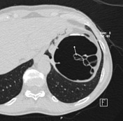
Radiographic Features
Depending on the degree of gastrointestinal involvement PCI may be diffuse, segmental or localized.
It is typically characterized by well-circumscribed cysts/”bubbles” within the walls of gastrointestinal structures. These cysts are surrounded by regular and non-inflamed mucosa.
Presence of small volume pneumoperitoneum.
If diffuse, it may appear to occupy large areas of the entire peritoneumabdomen with a honeycomb pattern that appears similar to that of interstitial lung disease.
Associations
Once identified is it important to try and identify an underlying cause, if present.
There have been proposed associations with chronic pulmonary, connective tissue and gastrointestinal diseases, including:
- Inflammatory Bowel Disease
- Necrotizing enterocolitis
- Sigmoid volvulus
- COPD
- Gastrointestinal Hypermobility
When the cysts rupture it may cause “benign pneumoperitoneum” as there is an absence of peritoneal irritation.
-<p><strong>Pneumatosis cystoides intestinalis</strong> (PCI) is a radiographic/descriptive entity characterized by the presence of multiple <em>gas filled cysts</em> in the submucosa and/or gastrointestinal subserosa of the small intestine. It is a subtype of pneumoatosis with specific features.</p><p>This process can occur anywhere along the gastrointestinal tract, including large bowel, rectum and mesentery. When associated with the colon it is known as pneumatosis cystoides coli (PCC).</p><p>Can be categorized as primary (idiopathic) or secondary.</p><p> </p><h4>Epidemiology</h4><p>PCI is an uncommon entity, reportedly occurring most commonly in the 6<sup>th</sup> decade, and has an incidence of ~0.03%.</p><p> </p><h4>Radiographic Features</h4><p>Depending on the degree of gastrointestinal involvement PCI may be diffuse, segmental or localized.</p><p>It is typically characterized by well-circumscribed cysts/”bubbles” within the walls of gastrointestinal structures. These cysts are surrounded by regular and non-inflamed mucosa.</p><p>Presence of small volume pneumoperitoneum.</p><p>If diffuse, it may appear to occupy the entire peritoneum with a honeycomb pattern that appears similar to that of interstitial lung disease.</p><p> </p><h4>Associations</h4><p>Once identified is it important to try and identify an underlying cause, if present.</p><p>There have been proposed associations with chronic pulmonary, connective tissue and gastrointestinal diseases, including:</p><ul>- +<p><strong>Pneumatosis cystoides intestinalis</strong> (PCI) is a radiographic/descriptive entity characterized by the presence of multiple <em>gas filled cysts</em> in the submucosa and/or gastrointestinal subserosa of the small intestine. It is a subtype of pneumatosis with specific features.</p><p>This process can occur anywhere along the gastrointestinal tract, including large bowel, rectum and mesentery. When associated with the colon it is known as pneumatosis cystoides coli (PCC).</p><p>Can be categorized as primary (idiopathic) or secondary.</p><h4>Epidemiology</h4><p>PCI is an uncommon entity, reportedly occurring most commonly in the 6<sup>th</sup> decade, and has an incidence of ~0.03%.</p><h4>Radiographic Features</h4><p>Depending on the degree of gastrointestinal involvement PCI may be diffuse, segmental or localized.</p><p>It is typically characterized by well-circumscribed cysts/”bubbles” within the walls of gastrointestinal structures. These cysts are surrounded by regular and non-inflamed mucosa.</p><p>Presence of small volume pneumoperitoneum.</p><p>If diffuse, it may appear to occupy large areas of the abdomen with a honeycomb pattern that appears similar to that of interstitial lung disease.</p><h4>Associations</h4><p>Once identified is it important to try and identify an underlying cause, if present.</p><p>There have been proposed associations with chronic pulmonary, connective tissue and gastrointestinal diseases, including:</p><ul>
References changed:
- 6. Rathi C, Pipaliya N, Poddar P, Pandey V, Ingle M, Sawant P. A Rare Case of Hypermobile Mesentery With Segmental Small Bowel Pneumatosis Cystoides Intestinalis. Intest Res. 2015;13(4):346. <a href="https://doi.org/10.5217/ir.2015.13.4.346">doi:10.5217/ir.2015.13.4.346</a> - <a href="https://www.ncbi.nlm.nih.gov/pubmed/26576141">Pubmed</a>
- 5. Schröpfer E & Meyer T. Surgical Aspects of Pneumatosis Cystoides Intestinalis: Two Case Reports. Cases J. 2009;2(1):6452. <a href="https://doi.org/10.4076/1757-1626-2-6452">doi:10.4076/1757-1626-2-6452</a> - <a href="https://www.ncbi.nlm.nih.gov/pubmed/19918585">Pubmed</a>
- 4. Arikanoglu Z. Pneumatosis Cystoides Intestinalis: A Single Center Experience. WJG. 2012;18(5):453. <a href="https://doi.org/10.3748/wjg.v18.i5.453">doi:10.3748/wjg.v18.i5.453</a> - <a href="https://www.ncbi.nlm.nih.gov/pubmed/22346251">Pubmed</a>
- 3. Ksiadzyna D & Peña A. Segmental Pneumatosis Cystoides Coli: Computed Tomography-Facilitated Diagnosis. Rev Esp Enferm Dig. 2015;108(8):510-3. <a href="https://doi.org/10.17235/reed.2015.3790/2015">doi:10.17235/reed.2015.3790/2015</a> - <a href="https://www.ncbi.nlm.nih.gov/pubmed/26652008">Pubmed</a>
- 2. Hosomi N, Yoshioka H, Kuroda C et al. Pneumatosis Cystoides Intestinalis: CT Findings. Abdom Imaging. 1994;19(2):137-9. <a href="https://doi.org/10.1007/bf00203487">doi:10.1007/bf00203487</a> - <a href="https://www.ncbi.nlm.nih.gov/pubmed/8199544">Pubmed</a>
- 1. Pohl J. Pneumatosis Cystoides Intestinalis. Video Journal and Encyclopedia of GI Endoscopy. 2013;1(2):393-4. <a href="https://doi.org/10.1016/s2212-0971(13)70174-2">doi:10.1016/s2212-0971(13)70174-2</a>
Systems changed:
- Gastrointestinal
Image 1 CT (lung window) ( create )

Image 2 CT (C+ portal venous phase) ( create )

Image 3 CT (lung window) ( create )


 Unable to process the form. Check for errors and try again.