
Sclerosing mesenteritis, also known as mesenteric panniculitis or retractile mesenteritis, is an uncommon idiopathic disorder characterized by chronic non-specific inflammation involving the adipose tissue of the bowel mesentery. It is often considered in the spectrum of autoimmune disease 21.
On this page:
Epidemiology
Typically this condition afflicts adults in their 60s with a mild male predilection, although reports vary 1,2,4,19.
Associations
Numerous associated conditions have been suggested including 1,4:
-
malignancy
systemic lymphoma 1,20 (up to 10% risk of developing lymphoma)
lung cancer 20
melanoma 20
colon cancer 20
renal cancer 20
myeloma 20
gastric carcinoma 4,20
recent abdominal surgery 4
-
systemic inflammatory conditions
autoimmune conditions (may be related to IgG4-related disease)
may be related to Weber-Christian disease 2
There is debate about the association between systemic inflammatory conditions and mesenteric panniculitis. Determining causation is difficult. The term "secondary mesenteric panniculitis" is reserved by some authors for patients with systemic inflammatory conditions. Most authors would not use the term when there is a local cause for mesenteric inflammation.
Clinical presentation
Clinical presentation can be variable with fever and abdominal pain common 20. Intestinal obstruction or ischemia, a mass, or diarrhea may also be present ref. Altered bowel habits and weight loss may be present in some cases ref. Occasionally, intermittent partial bowel obstruction is encountered 2. A firm left upper quadrant / central abdominal mass may be felt 2. In some situations sclerosing mesenteritis is asymptomatic.
Pathology
The small bowel mesentery is affected in most cases although the sigmoid mesocolon and omentum can also occasionally be involved.
The disease is said to pass through three stages, although some authors believe these to be separate entities 4:
mesenteric lipodystrophy: degeneration of mesenteric fat
mesenteric panniculitis: inflammatory reaction
retractile mesenteritis / sclerosing mesenteritis: fibrosis, which may be associated with distortion or lymphatic obstruction
Macroscopic appearance
Macroscopically, the mesentery is thickened with either solitary or multiple focal masses 4.
Microscopic appearance
Histology demonstrates 1:
lipid-laden macrophages (mesenteric lipodystrophy)
lymphocytic aggregates and lymphoid follicles (mesenteric panniculitis)
variable amounts of fibrosis (retractile mesenteritis)
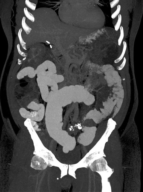
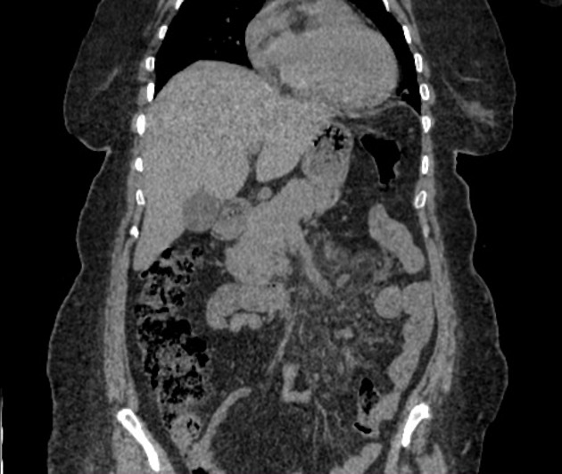
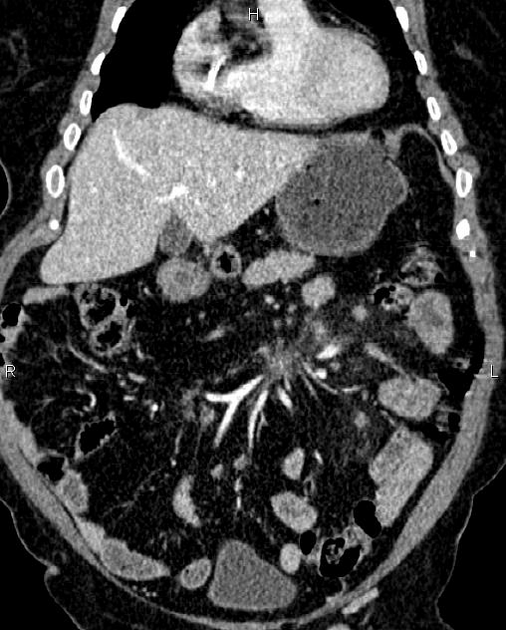
Radiographic features
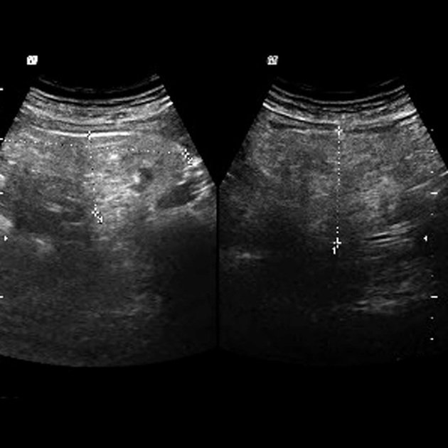
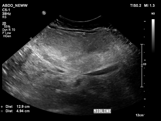
Ultrasound
Ultrasound typically demonstrates distortion and thickening of the root of the mesentery with a slight increase in echogenicity. Mass effect may be evident 3. A halo of sparing around vessels may be also seen on ultrasound as a region of hypoechoic fat 3. Color Doppler may show non-deviated mesenteric vessels within the mass 17.
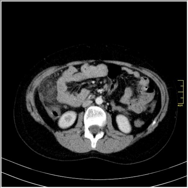
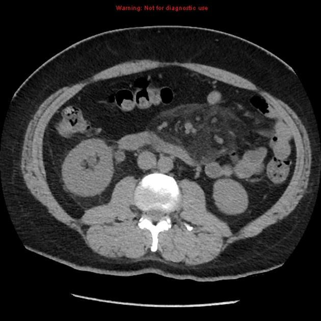
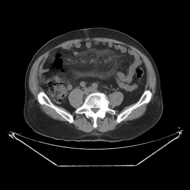
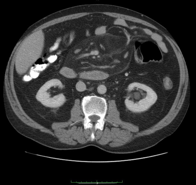
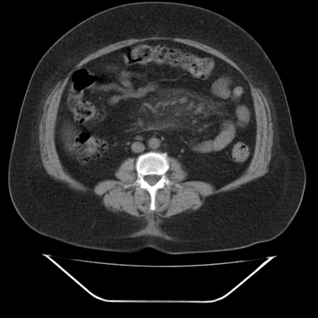
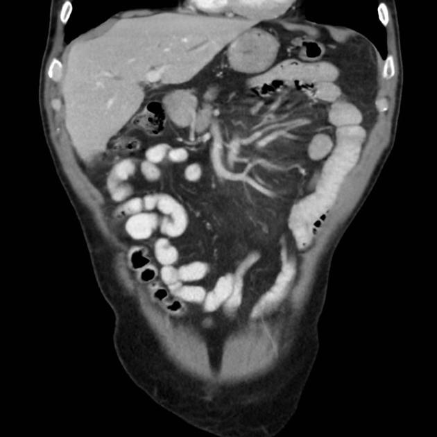
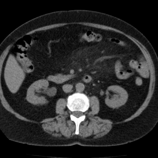
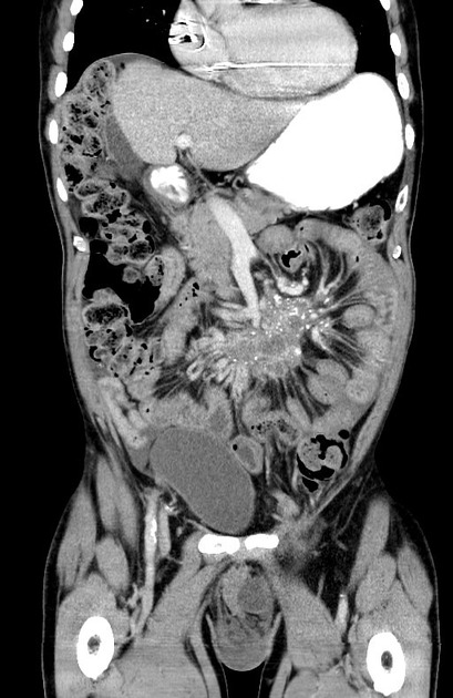
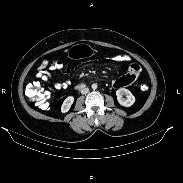
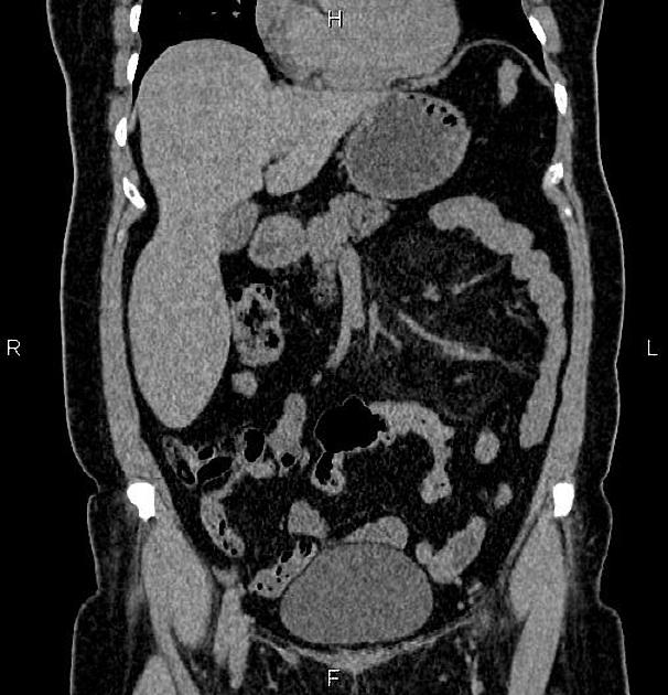
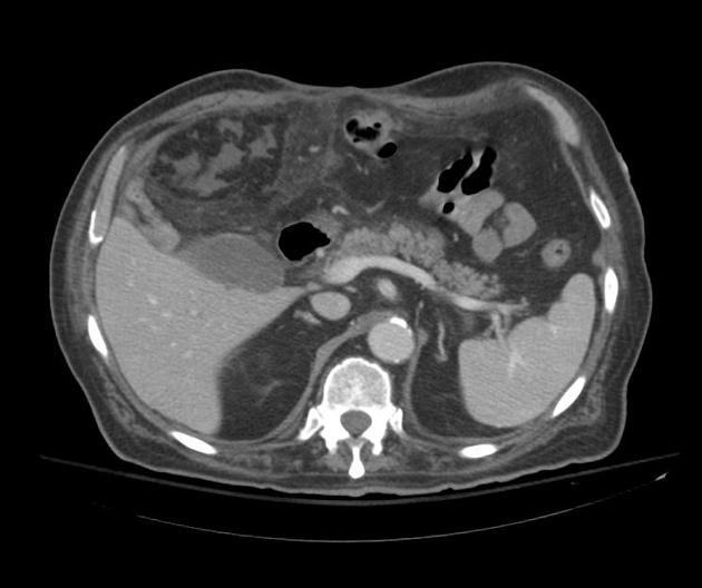
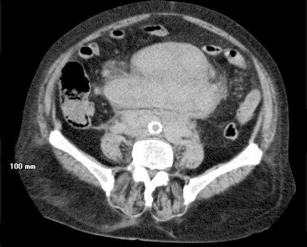
CT
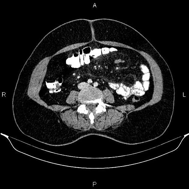
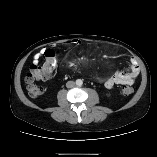
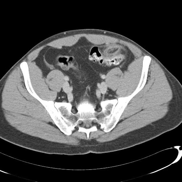
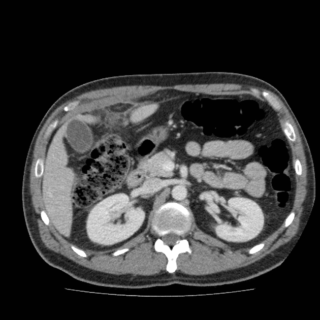
CT features are somewhat dependent on the main tissue component and include:
well-demarcated or ill-defined mesenteric mass-like lesion with surrounding "misty" attenuation
"misty" soft-tissue attenuation
The mesentery demonstrates mass effect and may have a ground glass opacity (misty mesentery). Typically the traversing mesenteric vessels and soft tissue nodules have a spared fat halo (this has sometimes been referred to as the fat ring sign). Its orientation is aligned with the root of the jejunal mesentery. Punctate/coarse calcifications (~20%), as well as small lymph nodes (usually <5 mm), may be present within the region.
The Coulier criteria can be used to diagnose sclerosing mesenteritis on CT when 3 of the following criteria are present 8:
mesenteric mass which defines and displaces nearby structures without invading them
inhomogenous fat density within the mass attenuates higher than adjacent retroperitoneal or meso-colonic fat
contains small soft tissue nodes, typically <10 mm
low attenuation fatty "halo sign" surrounds lymph nodes or mesenteric vessels
hyperattenuating pseudocapsule may surround the affected area in the absence of ascites or known malignancy involving the mesentery
The last two signs are considered inconstant but very specific.
MRI
Findings are similar to CT. One report described a fibrous capsule around the inflammation 12.
Nuclear medicine
FDG PET-CT is useful for differentiating between sclerosing mesenteritis and malignant mesenteric involvement.
sclerosing mesenteritis (or one of its stages): not FDG-avid
malignant mesenteric involvement: FDG-avid
This is especially useful in patients with lymphoma 10. If in doubt, biopsy may be indicated in selected patients, even in asymptomatic lesions 11,19.
Treatment and prognosis
The mainstay of treatment is supportive, as the disease is typically self-limiting 19. If severe or protracted, medical therapy with corticosteroids, cyclophosphamide or azathioprine can be contemplated 2. Mesenteric panniculitis cannot be completely resected and surgery is of no benefit.
While local lymphoma (in up to 15% of cases) 2,4, and a more general association with malignancy of 37-56% have been suggested 16,24, but a true association has not been proved. In patients diagnosed with mesenteric panniculitis, lymphadenopathy >12 mm, and absence of a 'fat halo' around lymph nodes and vessels increased the risk of subsequent malignancy diagnosis in one study 24. Limitations of studies suggesting a malignancy association have been highlighted, and contrary evidence including a systematic review and meta-analysis, and case matched cohort studies have dismissed such an association 25-27.
History and etymology
It was first described by Jura in 1924 as “retractile mesenteritis” and further labeled as “mesenteric panniculitis” by Odgen later in the 1960s.
Differential diagnosis
Mesenteric panniculitis cannot be diagnosed on imaging without excluding many other possible causes of a misty mesentery 22.
General imaging differential considerations include:
mesenteric carcinoid tumor
-
mesenteric lymphadenopathy due to malignancy:
metastatic disease
-
mesenteric lymphadenopathy due to inflammation:
systemic illness
mesenteric lipoma: especially if large and if there are lymph nodes within it


 Unable to process the form. Check for errors and try again.