
Skull (submentovertex view)
Updates to Article Attributes
Body
was changed:
The skull submentovertex view is an angled inferosuperior radiograph of the base of skull. As this view involves radiographic positioning that is uncomfortable for the patient and with CT being more sensitive to bony detail, this view is rapidly becoming obsolete.
Indications
This view is useful in assessing potential pathology from trauma or disease progression to the basal skull structures 1-4, including the foramen ovale, foramen spinosum and sphenoid sinuses.
It is imperative that any cervical spine subluxations or fractures on acute trauma patients is excluded before proceeding with this view.
Patient position
- depending on how the patient presents
:- erect: patient leans back on a chair with back support, facing away from the upright bucky
- supine: elevate the shoulders using a firm pillow, allowing the head to tilt backwards
- the neck is hyperextended until
- the infraorbitomeatal line (IOML) is parallel with the receptor
- the skull vertex is in contact with the center of the receptor
- ensure the midsagittal plane (MSP) is perpendicular to the receptor
Technical factors
- inferosuperior projection
-
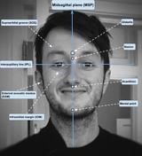
centring point
- 4 cm inferior to the mandibular mental point (see Figure 2)
- beam exits at skull vertex
-
collimation
- anterior to include mandibular mentum
- posterior to include occipital bone
- lateral to include the skin margin
-
orientation
- portrait
-
detector size
- 24 cm x 30 cm
-
exposure
- 75-80 kVp
- 20-25 mAs
-
SID
- 100 cm
-
grid
- yes (this can vary departmentally)
Image technical evaluation
- the mandibular mentum should be demonstrated just slightly anterior to the ethmoid sinuses
- if too far anterior to the ethmoid sinuses, depress the patient's chin or angle more caudal
- if demonstrated posterior to the ethmoid sinuses, further extend the neck or angle more cephalic
- there should be equal distance between the mandibular rami and the lateral cranial cortex
- i.e. an increased mandibular ramus-lateral cranial cortex distance on the right side means the patient's head was tilted towards the right
Practical points
- patients who are supine for this view may become dizzy after a few minutes due to an increased intracranial pressure 4; the erect method may prevent this
- learning your skull anatomy and positioning lines makes reading positioning guides a whole lot easier
- placing a physical side marker can be useful in determining which side is which
-<p>The <strong>skull submentovertex view</strong> is an angled inferosuperior radiograph of the base of skull. As this view involves radiographic positioning that is uncomfortable for the patient and with CT being more sensitive to bony detail, this view is rapidly becoming obsolete.</p><h4>Indications</h4><p>This view is useful in assessing potential pathology from trauma or disease progression to the basal skull structures <sup>1-4</sup>, including the <a title="Foramen ovale (skull)" href="/articles/foramen-ovale-skull">foramen ovale</a>, <a title="Foramen spinosum" href="/articles/foramen-spinosum">foramen spinosum</a> and sphenoid sinuses.</p><p>It is imperative that any cervical spine subluxations or fractures on acute trauma patients is excluded before proceeding with this view.</p><h4>Patient position</h4><ul>-<li>depending on how the patient presents:<ul>- +<p>The <strong>skull submentovertex view</strong> is an angled inferosuperior radiograph of the base of skull. As this view involves radiographic positioning that is uncomfortable for the patient and with CT being more sensitive to bony detail, this view is rapidly becoming obsolete.</p><h4>Indications</h4><p>This view is useful in assessing potential pathology from trauma or disease progression to the basal skull structures <sup>1-4</sup>, including the <a href="/articles/foramen-ovale-skull">foramen ovale</a>, <a href="/articles/foramen-spinosum">foramen spinosum</a> and sphenoid sinuses.</p><p>It is imperative that any cervical spine subluxations or fractures on acute trauma patients is excluded before proceeding with this view.</p><h4>Patient position</h4><ul>
- +<li>depending on how the patient presents<ul>
References changed:
- 1. John Lampignano, Leslie E. Kendrick. Bontrager's Textbook of Radiographic Positioning and Related Anatomy. (2017) <a href="https://books.google.co.uk/books?vid=ISBN9780323399661">ISBN: 9780323399661</a><span class="ref_v4"></span>
- 2. A. Stewart Whitley, Charles Sloane, Graham Hoadley, Adrian D. Moore. Clark's Positioning in Radiography 12Ed. (2005) <a href="https://books.google.co.uk/books?vid=ISBN9780340763902">ISBN: 9780340763902</a><span class="ref_v4"></span>
- 3. Kathy McQuillen Martensen. Radiographic Image Analysis. (2014) <a href="https://books.google.co.uk/books?vid=ISBN9780323280525">ISBN: 9780323280525</a><span class="ref_v4"></span>
- 4. Eugene D. Frank, Bruce W. Long, Barbara J. Smith, Vinita Merrill. Merrill's Atlas of Radiographic Positioning and Procedures. (2020) <a href="https://books.google.co.uk/books?vid=ISBN9780323073219">ISBN: 9780323073219</a><span class="ref_v4"></span>
Sections changed:
- Radiography
Systems changed:
- Central Nervous System
- Head & Neck
Images Changes:
Image 1 Diagram ( create )

Image 2 Annotated image (annotated frontal facial photograph) ( create )


 Unable to process the form. Check for errors and try again.