
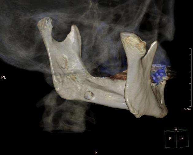
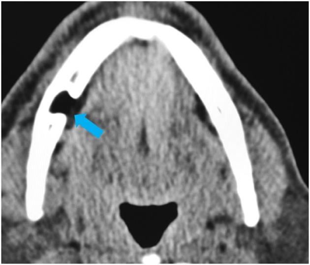
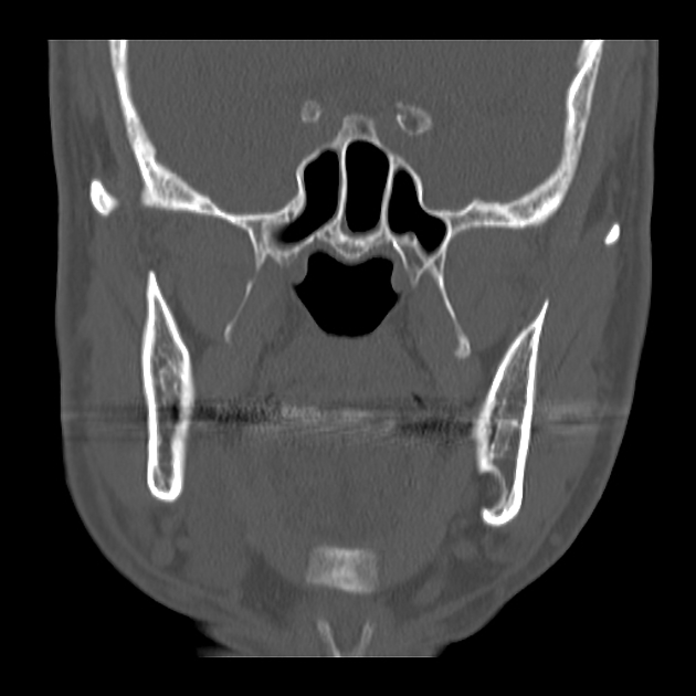
A Stafne bone cavity is a cortical defect near the angle of the mandible below the mandibular canal. It is usually an incidental finding and represents a depression in the medial aspect of the mandible filled by part of the submandibular gland or adjacent fat.
On this page:
Terminology
A Stafne bone cavity is also known as a Stafne defect, Stafne bone cyst, static bone cavity of the mandible, or lingual salivary gland inclusion defect. Strictly speaking, it is not a cyst since it does not contain any fluid. Therefore, the term "Stafne bone cavity" is preferred 2.
Epidemiology
Stafne bone cavities are most frequently seen in middle-aged men. The estimated prevalence ranges around 0.10-0.48% 2.
Pathology
Stafne bone cavities are thought to result from remodeling of the bone by adjacent salivary tissue and have been noted to regress following resection of the gland nearby.
Location
It generally occurs in the area between the mandibular first molar and the mandibular angle 6.
Radiographic features
The Stafne bone cavity tends to not increase in size or change in radiographic appearance over time (hence the term "static bone cyst"), and this can be used to help confirm the diagnosis.
Plain radiograph
Stafne bone cavities are usually discovered incidentally during routine dental radiography.
Radiographically, it is a well-circumscribed, unilocular, round, radiolucent defect, 1-3 cm in size, usually between the inferior alveolar nerve and the inferior border of the posterior mandible between the molars and the angle of the jaw. The radiolucent defect may be superimposed on the lower anterior teeth and be mistaken for an odontogenic lesion. Sometimes the defect may interrupt the contour of the lower border of the mandible and may be palpable.
Fluoroscopy
Sialography may be sometimes used to help demonstrate the salivary gland tissue within the bone.
CT
CT will show a shallow defect through the medial cortex of the mandible with a corticated rim and no soft tissue abnormalities, with the exception of a portion of the submandibular gland herniating into the defect.
MRI
MRI can delineate the continuation of the submandibular gland into the mandibular defect as an alternative to CT.
Differential diagnosis
It should not be confused with other lytic lesions of the jaw.
History and etymology
It is named after Edward C Stafne, an American dentist (1894-1981).


 Unable to process the form. Check for errors and try again.