
Tuberculous meningitis is the most common presentation of intracranial tuberculosis, and usually refers to infection of the leptomeninges. Uncommonly tuberculosis can be limited to the pachymeninges (dura mater), it is called tuberculous pachymeningitis and is discussed separately.
The remainder of this article pertains to leptomeningeal tuberculosis, which involves the arachnoid mater and pia mater.
On this page:
Epidemiology
Tuberculous meningitis, although seen in all age groups, has a peak incidence in childhood (particularly 0-4 years of age) in high prevalence areas. In low prevalence areas, it is more frequently encountered in adolescents and adults.
Important risk factors include:
Clinical presentation
Low-grade fever with headache is a prodromal manifestation. Most common clinical manifestations are fever, headache, vomiting and neck stiffness. Cranial nerve palsies of 3rd, 4th and 6th nerves may be seen. Seizures, focal neurological deficits, stupor and coma may be seen in late stages.
CSF analysis reveals lymphocytosis, increased protein level and decreased glucose levels.
Pathology
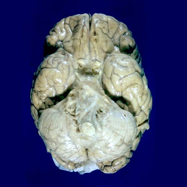
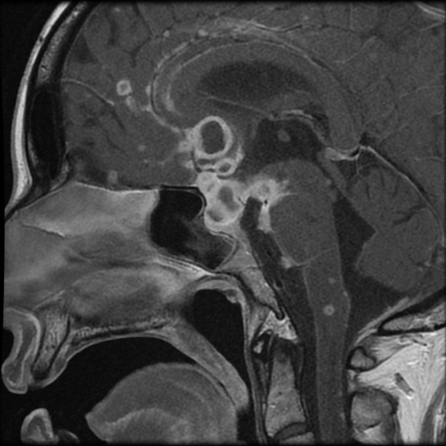
Tuberculous meningitis is caused by Mycobacterium tuberculosis. The infection spreads hematogenously from a distant focal point, usually pulmonary tuberculosis and lodges immediately deep to the pia forming Rich foci. These can rupture into the subarachnoid space, forming an exudate. This purulent material is primarily located in the vicinity of basal cisterns: inferomedial surface of frontal lobe, anteromedial surface of temporal lobes, superior cerebellum and floor of the fourth ventricle.
From here, infection spreads to interpeduncular cisterns, around optic chiasm and to pontomesencephalic, ambient and suprasellar cisterns. Although the exudate can reach the Sylvian fissures it uncommonly extends over the cerebral convexities 3.
Choroid plexitis may also be a late manifestation as is mass-like regions of caseous necrosis within this exudate.
Radiographic features
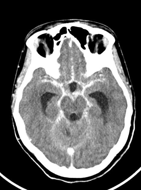
CT
non-contrast scans may be normal
-
later complications may be visible including:
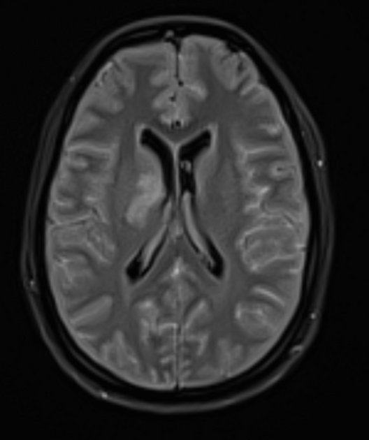
infarcts due to arteritis (especially in children)
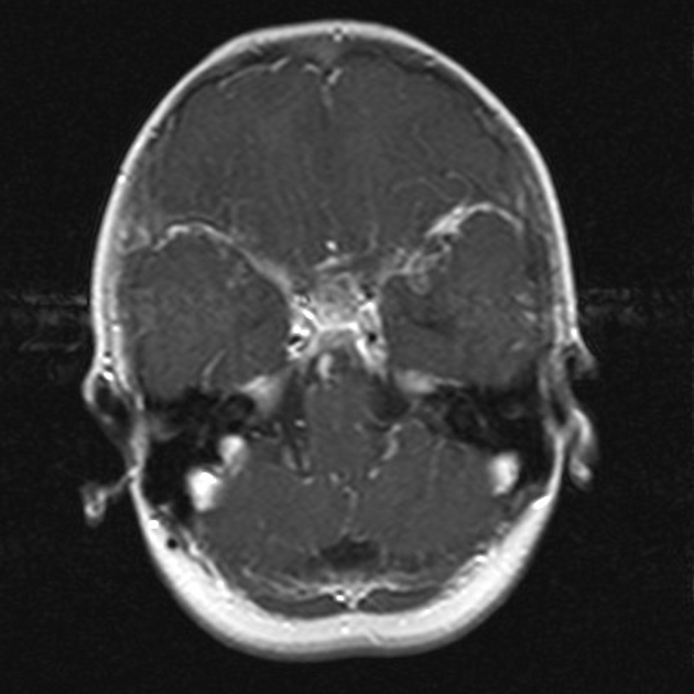
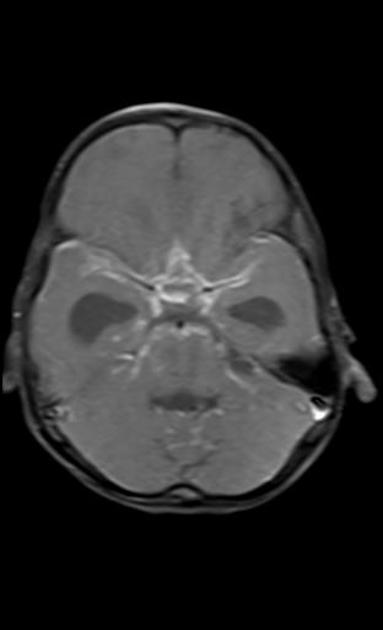
Following contrast administration a number of additional features may be visible:
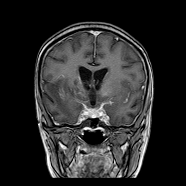
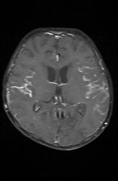
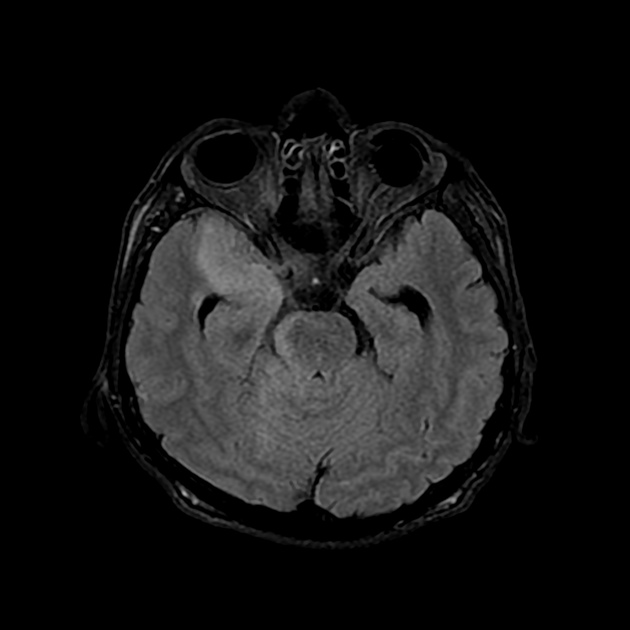
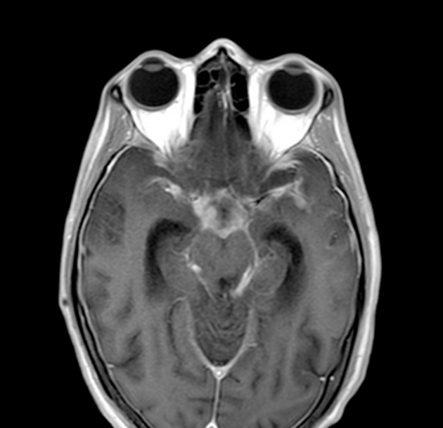
basal enhancing exudates
leptomeningeal enhancement, along sylvian fissures, tentorium uncommonly convexities
ependymitis may be present
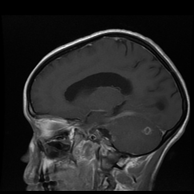
MRI
-
T1
normal initially
T1 shortening may be seen after progression of disease 3
-
T2
normal initially
T2 shortening is seen after disease progression 3
T1 C+ (Gd): diffuse basal enhancement with enhancing exudates
magnetization transfer (MT) spin echo: significantly lower MT ratio is seen in tuberculous meningitis as compared to fungal and pyogenic meningitis
Treatment and prognosis
Anti-tuberculosis regimen is started after confirmation of diagnosis. Treatment of complications (e.g. drainage of hydrocephalus) is also performed.
Complications
-
arteritis that may result in ischemic infarcts 5
affects one-third of cases
more common in children
cranial neuropathies: most affected nerves are 3rd, 4th and 6th nerves
Differential diagnosis
General imaging differential considerations include:


 Unable to process the form. Check for errors and try again.