
Volar intercalated segment instability
Updates to Article Attributes
Volar intercalated segment instability (VISI) is a type of carpal instability featuring volar tilt of the lunate. It is less often encountered than dorsal intercalated segment instability (DISI).
Clinical presentation
It presents in most cases with nonspecific wrist pain and a "clunking" on the ulnar deviation of the wrist.
Pathology
VISI can occur because of a disruption of radiocarpal ligaments on the ulnar side of the wrist. The main ligaments involved in this instability are thought to be the ulnar half of the volar arcuate ligament 6 and the lunotriquetral ligament 6,7. It may be static or dynamic.
A VISI alignment can be found in uninjured wrists and therefore considered a normal variant for patients with lax ligaments.
Radiographic features
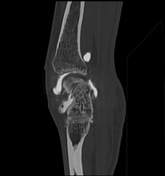
The main radiographic features are a volar rotation of the lunate and dorsal rotation of the capitate and hamate.
Plain radiograph
Abnormal carpal angles are seen on any lateral or sagittal imaging of the wrist when the wrist is in a neutral position:
- scapholunate angle <30°
- capitolunate angle >30°
Treatment and prognosis
The mainstay of treatment is surgery, either by early reduction and casting with K-wires or by capitolunate fusion.
See also
-<p><strong>Volar intercalated segment instability</strong> (<strong>VISI</strong>) is a type of <a title="Carpal instability" href="/articles/carpal-instability">carpal instability</a> featuring <a href="/articles/anatomical-position">volar</a> tilt of the lunate. It is less often encountered than <a href="/articles/dorsal-intercalated-segment-instability">dorsal intercalated segment instability</a> (DISI).</p><h4>Clinical presentation</h4><p>It presents in most cases with nonspecific wrist pain and a "clunking" on the ulnar deviation of the wrist.</p><h4>Pathology</h4><p>VISI can occur because of a disruption of radiocarpal ligaments on the ulnar side of the wrist. The main ligaments involved in this instability are thought to be the ulnar half of the volar arcuate ligament <sup>6 </sup>and the <a href="/articles/lunotriquetral-ligament">lunotriquetral ligament</a> <sup>6,7</sup>. It may be static or dynamic.</p><p>A VISI alignment can be found in uninjured wrists and therefore considered a normal variant for patients with lax ligaments.</p><h4>Radiographic features</h4><p>The main radiographic features are a volar rotation of the lunate and dorsal rotation of the capitate and hamate.</p><h5>Plain radiograph</h5><p>Abnormal carpal angles are seen on any lateral or sagittal imaging of the wrist when the wrist is in a neutral position:</p><ul>- +<p><strong>Volar intercalated segment instability</strong> (<strong>VISI</strong>) is a type of <a href="/articles/carpal-instability">carpal instability</a> featuring <a href="/articles/anatomical-position">volar</a> tilt of the lunate. It is less often encountered than <a href="/articles/dorsal-intercalated-segment-instability">dorsal intercalated segment instability</a> (DISI).</p><h4>Clinical presentation</h4><p>It presents in most cases with nonspecific wrist pain and a "clunking" on the ulnar deviation of the wrist.</p><h4>Pathology</h4><p>VISI can occur because of a disruption of radiocarpal ligaments on the ulnar side of the wrist. The main ligaments involved in this instability are thought to be the ulnar half of the volar arcuate ligament <sup>6 </sup>and the <a href="/articles/lunotriquetral-ligament">lunotriquetral ligament</a> <sup>6,7</sup>. It may be static or dynamic.</p><p>A VISI alignment can be found in uninjured wrists and therefore considered a normal variant for patients with lax ligaments.</p><h4>Radiographic features</h4><p>The main radiographic features are a volar rotation of the lunate and dorsal rotation of the capitate and hamate.</p><h5>Plain radiograph</h5><p>Abnormal carpal angles are seen on any lateral or sagittal imaging of the wrist when the wrist is in a neutral position:</p><ul>
Image 2 ( create )

Image 3 ( create )

Image 4 Diagram (lateral) ( update )
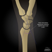
Image 5 ( create )

Image 6 Diagram (lateral) ( update )
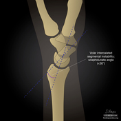
Image 7 X-ray (Lateral) ( update )

Image 8 CT (bone window) ( update )


 Unable to process the form. Check for errors and try again.