
Atypical parathyroid adenoma
Updates to Study Attributes
Macroscopic and Microscopic Findings
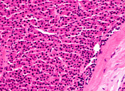
The specimen consists of a 1.20g, 12 mm well circumscribed tumour with peripheral fibrous capsule, composed of solid sheets of parathyroid epithelial oncocytic cells showing a vaguely organoid architecture without interspersed fat and showing a scant amount of peripheral normal appearing parathyroid gland. Tumour cells are medium in size with rounded to ovoid nuclei and a moderate amount of oncocytic cytoplasm. Thick, hyalinised fibrous stromal band traverse the lesion. There is no tumour cell necrosis or haemorrhage. There is no significant mitotic activity.
20x H&E: Low-power view shows sheets of parathyroid cells with a peripheral fibrous capsule and traversing hyalinised fibrous bands. Normal surrounding parenchymal tissue is not seen in this section.
40x H&E: Medium-power view showing hyalinised bands, with epithelial cells displaying acinar architectural patterns which are subtly visible at this power.
100x H&E: Closer view.
200x H&E: This high power view highlights the bland, monomorphous and mitotically inactive appearance of the parathyroid cells.
Immunohistochemistry
A galectin-3 (Gal-3) immunohistochemical stain was performed, which was negative.
Image Pathology (H&E) ( update )

Image Pathology (H&E) ( update )

Image Pathology (H&E) ( update )
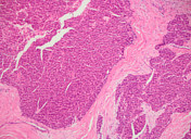
Image Pathology (H&E) ( update )


 Unable to process the form. Check for errors and try again.