
Brown tumours - hyperparathyroidism
- Radiopaedia Events Pty Ltd, Speaker fees (past)
- Integral Diagnostics, Shareholder (ongoing)
- Micro-X Ltd, Shareholder (ongoing)
Updates to Case Attributes
The case illustrates the radiological features of hyperparathyroidism including giant cell tumours at the iliac bones, osteomalacia of the examined bones, stress fractures of the inferior pubic rami, bilateral erosive changes of the articular surfaces of both sacroiliac joints, subperiosteal resorption of the medial aspect of the left femoral neck and erosive changes of the medial sternal heads.
99mTc sestamibi (99mTc-MIBI) imaging gave a false negative study and failed to localise the parathyroid adenoma.
However, CT and MRI of the neck showed an irregular-shaped lesion at the suprasternal region showing mixed fatty and solid texture.
The patient later underwent surgical resection of the suprasternal parathyroid tumour and the pathological diagnosis was parathyroid adenoma.
False negative 99mTc sestamibi results might be secondary to small tumour size, cystic components, multiglandular disease, and the site of parathyroid adenoma. In this case, other imaging modalities such as 4D CT scan, US, MRI, or even venous sampling might help to localise parathyroid adenomas.
-<p>The case illustrates the radiological features of hyperparathyroidism including giant cell tumours at the iliac bones, osteomalacia of the examined bones, stress fractures of the inferior pubic rami, bilateral erosive changes of the articular surfaces of both sacroiliac joints, subperiosteal resorption of the medial aspect of the left femoral neck and erosive changes of the medial sternal heads.</p><p>99mTc sestamibi (99mTc-MIBI) imaging gave a false negative study and failed to localise the parathyroid adenoma.</p><p>However, CT and MRI of the neck showed an irregular-shaped lesion at the suprasternal region showing mixed fatty and solid texture.</p><p>The patient later underwent surgical resection of the suprasternal parathyroid tumour and the pathological diagnosis was parathyroid adenoma.</p><p>False negative 99mTc sestamibi results might be secondary to small tumour size, cystic components, multiglandular disease, and the site of parathyroid adenoma. In this case, other imaging modalities such as 4D CT scan, US, MRI, or even venous sampling might help to localise parathyroid adenomas.</p>- +<p>The case illustrates the radiological features of hyperparathyroidism including giant cell tumours at the iliac bones, osteomalacia of the examined bones, stress fractures of the inferior pubic rami, bilateral erosive changes of the articular surfaces of both sacroiliac joints, subperiosteal resorption of the medial aspect of the left femoral neck and erosive changes of the medial sternal heads.</p><p>99mTc sestamibi (99mTc-MIBI) imaging gave a false negative study and failed to localise the parathyroid adenoma.</p><p>However, CT and MRI of the neck showed an irregular-shaped lesion at the suprasternal region showing mixed fatty and solid texture.</p><p>The patient later underwent surgical resection of the suprasternal parathyroid tumour and the pathological diagnosis was parathyroid adenoma.</p><p>False negative 99mTc sestamibi results might be secondary to small tumour size, cystic components, multiglandular disease, and the site of parathyroid adenoma. In this case, other imaging modalities such as 4D CT, US, MRI, or even venous sampling might help to localise parathyroid adenomas.</p>
Systems changed:
- Paediatrics
Tags changed:
- endocrinesurgery
- endocrine surgery
- endocrine
Updates to Link Attributes
Updates to Primarylink Attributes
Updates to Study Attributes
An expansile altered marrow signal of the left superior pubic body and superior pubic ramus eliciting low T2 with tiny cystic hyperintense foci of high signal. It is seen associated with markedly thinned out cortex and mild cortical erosions, yet no extra-osseous soft tissue components.
A similar smaller lesion is seen implicating the right inferior pubic ramus.
Bilateral inferior pubic ramus stress fractures and surrounding edema.
*TheThe characteristic Lowlow T2 signal and tiny internal cysts assas well as the bilateral inferior pubic rami stress fractures were both suggestive of hyperparathyroidism and brown tumors*.
Updates to Study Attributes
osteomalacia of the examined bones
Left pubic and smaller right iliac and right inferior pubic ramus lytic lesions (likely brown tumors)
bilateral inferior pubic rami stress fractures
bilateral erosive changes of the articular surfaces of both sacroiliac joints
subperiosteal resorption of the medial aspect of the left femoral neck
CT findings confirmed the previous possibility of hyperparathyroidism and brown tumors.
Updates to Study Attributes
Normal Parathyroidparathyroid scan. No evidence of parathyroid adenoma.
Updates to Study Attributes
An irregular-shaped soft tissue lesion is seen at the suprasternal region abutting the aortic arch and the brachiocephalic artery, probably representing ectopic parathyroid adenoma.
Image MRI (T2) ( update )
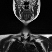
Image MRI (T2) ( update )
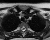
Image 1 MRI (T2) ( update )
Image 2 MRI (T2) ( update )


 Unable to process the form. Check for errors and try again.