
Gluteal atrophy
Diagnosis certain
Disclosures
- updated 26 Jan 2024:
Nothing to disclose
Updates to Study Attributes
Findings
was changed:
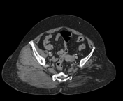
Left-sided gluteal muscles are markedmarkedly atrophic in size with relativelyrelative sparing of the gluteus maximus. An imaged portion of the left upper thigh also shows atrophy of the anterior and posterior muscular compartments however,, though the sartoris muscle appears spared.Fatty replacementof the above-mentioned muscles is seen.
The right side gluteal region and imaged thigh are unremarkable.
Visualized bones are normal.
Images Changes:
Image 33 CT (C+ portal venous phase) ( create )
Annotation 17623
changed from ,0 arrows,0 labels to atrophy,1 arrow,1 label.
Updates to Case Attributes
Body
was changed:
CT features are left-sided gluteal muscle atrophy however, no. No history of a definite cause was identified, such as poliomyelitis is provided which could. This finding may be duesecondary to long-standing disuse.
Co-contributor: Dr,. Anwar-ul-Haq Zadran.
-<p>CT features are left-sided gluteal muscle atrophy however, no history of definite cause such as poliomyelitis is provided which could be due to long-standing disuse.</p><p>Co-contributor: Dr, Anwar-ul-Haq Zadran.</p><p></p><p></p>- +<p>CT features are left-sided gluteal muscle atrophy. No history of a definite cause was identified, such as poliomyelitis. This finding may be secondary to long-standing disuse.</p><p>Co-contributor: Dr. Anwar-ul-Haq Zadran</p><p></p>


 Unable to process the form. Check for errors and try again.