
Hemangioblastoma
Diagnosis almost certain
Updates to Case Attributes
Diagnostic Certainty
was set to
.
Presentation
was changed:
26 years old man with a history of progressive headaches.
Age
was set to
26.
Gender
was set to
Male.
Body
was changed:
The patient was submitted to total surgical removal that confirms the diagnosis of haemangioblastoma. 8 months after the surgery, a control MRI exams showed signs of relapse.
-<p>The patient was submitted to total surgical removal that confirms the diagnosis of <a href="/articles/haemangioblastoma-cns" title="Hemangioblastoma">hemangioblastoma</a>. 8 months after the surgery, a control MRI exams showed signs of relapse.</p>- +<p>The patient was submitted to total surgical removal that confirms the diagnosis of <a href="/articles/haemangioblastoma-central-nervous-system-1">haemangioblastoma</a>. 8 months after the surgery, a control MRI exams showed signs of relapse.</p>
Updates to Study Attributes
Modality
was set to
MRI.
Caption
was added:
MRI Brain (8 months later)
Findings
was added:
A solid contrast enhanced nodule is identified within the inferior portion of the 4th ventricle and in keeping with tumour recurrence.
Updates to Study Attributes
Caption
was added:
MRI Brain
Findings
was added:
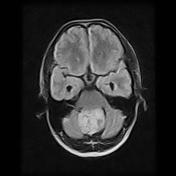
There is a mid-line solid cerebellar mass compressing the 4th ventricle posteriorly and promoting hydrocephalus. The mas show vivid contrast enhancement and small flow voids on T2.
Images Changes:
Image MRI (FLAIR) ( update )

Stack
was set to
.
Single Or Stack Root
was set to
.
Image MRI (FLAIR) ( update )
Stack
was set to
.
Single Or Stack Root
was set to
.
Perspective
was set to
Axial.
Specifics
changed from T1 to FLAIR.
Image MRI (T1) ( update )

Perspective
was set to
Sagittal.
Image MRI (T2) ( update )

Perspective
was set to
Axial.
Specifics
changed from to T2.
Image MRI (T1 C+) ( update )

Perspective
was set to
Axial.
Image MRI (T1 C+) ( update )

Perspective
was set to
Sagittal.


 Unable to process the form. Check for errors and try again.