
Hemothorax
Updates to Study Attributes
Bilateral haemothorax, right greater than left, accounting for the density difference seen on the supine chest x-ray. Note the haematocrit level within the right haemothorax. Mild atelectasis is seen adjacent to the haemothoraces and likely small volume pulmonary contusion in the right lung base where a small traumatic pneumatocele is seen. Subtle inward angulation of multiple anterobasal ribs are consistent with incomplete fractures.A few tiny loculesright T10 transverse process fracture was better appreciated on sagittal images and a fracture of the right superior aspect of the T10 vertebral body. Locules of soft-tissue gas are seen innear the soft tissues in the region ofT10 injury and also near the rib fractures and trace. Trace bilateral pneumothorax is seen anterobasally.
Image CT (bone window) ( update )
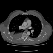
Image 3 CT (bone window) ( create )
Updates to Case Attributes
This case is a nice example of a supine haemothorax on a trauma chest radiograph being identifiable as a generalised increase in hemithorax density compared to the contralateral side.
CT Findings
- Bilateral haemothorax, right greater than left
haematocrit level within the right haemothorax- mild atelectasis adjacent to the haemothoraces
- small pulmonary contusion and traumatic pneumatocele right lung base
- multiple bilateral anterobasal incomplete rib fractures
-
a few tinyright T10 transverse process and vertebral body fracture with adjacent locules of gasin the soft tissues adjacent to rib fractures - trace bilateral pneumothorax anterobasally (very tiny!)
-<li>haematocrit level within the right haemothorax</li>-<li>a few tiny locules of gas in the soft tissues adjacent to rib fractures</li>- +<li>right T10 transverse process and vertebral body fracture with adjacent locules of gas</li>


 Unable to process the form. Check for errors and try again.