
Hip arthrogram injection (fluoroscopic guided)
Diagnosis not applicable
Updates to Study Attributes
Modality
changed from to Fluoroscopy.
Caption
was added:
Arthrogram injection pre MRI
Findings
was added:
Arthrogram solution containing iodinated contrast distends the hip joint. The needle has been removed.
Images Changes:
Image 1 Fluoroscopy (Frontal) ( create )

Updates to Study Attributes
Caption
was added:
Arthrogram injection pre MRI
Findings
was changed:
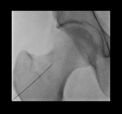
Technically successful right hip fluoroscopic-guided arthrogram injection. Contrast flows freely and distends head neck aspect ofinto the hip joint at the level of the head-neck junction. An oblique needle approach used with a 22G 90mm Quincke needle.
Images Changes:
Image Fluoroscopy (Frontal) ( update )

Stack
was set to
.
Single Or Stack Root
was set to
.
Image Fluoroscopy (Frontal) ( update )
Stack
was set to
.
Single Or Stack Root
was set to
.
Perspective
was set to
Frontal.
Image 1 Fluoroscopy (Frontal) ( create )
Updates to Study Attributes
Findings
was removed:
An oblique needle approach used with a 22G 90mm Quincke needle. Iodinated contrast confirms an intra-articular needle tip position.
Caption
was added:
Modality
was removed.
Images Changes:
Image ( destroy )

Size
was set to
.
Width
was set to
.
Height
was set to
.
Filename
was removed.
Position
was set to
.
Stack
was set to
.
Specifics
was set to
.
Description
was added:
Perspective
was removed.
Content Type
was removed.
Contributor
was set to
.
Thumbnail Files
was removed.
Original Filename
was set to
.
Single Or Stack Root
was set to
.
Updates to Quizquestion Attributes
Answer
was changed:
Yes. The needle tip is within the joint and contrast extends to surround the superior head-neck junction of the hip joint.


 Unable to process the form. Check for errors and try again.