
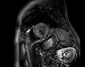
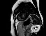
Hypertrophic cardiomyopathy - apical pattern with midventricular obstruction
Diagnosis certain
Updates to Study Attributes
Images Changes:
Image MRI (LGE with look locker) ( update )
Perspective
was set to
Small axis.
Specifics
was set to
LGE with look locker.
Image MRI (PSIR gado) ( update )
Perspective
was set to
Small axis.
Specifics
was set to
PSIR gado.
Image MRI (PSIR gado) ( update )

Perspective
was set to
Long Axis.
Specifics
was set to
PSIR gado.
Image MRI (PSIR gado) ( update )

Perspective
was set to
4 chamber.
Specifics
was set to
PSIR gado.
Image MRI (T1 mapping) ( update )
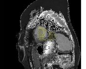
Perspective
was set to
Small axis.
Specifics
was set to
T1 mapping.
Image 5 MRI (T1 mapping) ( create )
Image 6 MRI (PSIR gado) ( create )
Image 7 MRI (LGE with look locker) ( create )
Image 8 MRI (PSIR gado) ( create )
Image 9 MRI (PSIR gado) ( create )


 Unable to process the form. Check for errors and try again.