

Interlobar loculated pneumothorax
Updates to Study Attributes
Chest X ray
The radiographic study of the chest, the anteroposterior projection, particularly the lateral one, shows an air-filled cystic lesion, delimited by thin and sclerotic walls, in the central part of the right lung. There are no other types of lesions or pleural effusion. The mediastinum is on the axis, the volume is within limits.
Image X-ray (Frontal) ( update )
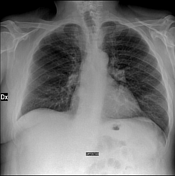
Image X-ray (Lateral) ( update )
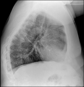
Image 1 X-ray (Frontal) ( destroy )
Image 1 X-ray (Lateral) ( destroy )
Image 1 X-ray (Lateral) ( destroy )
Image 1 X-ray (Lateral) ( destroy )
Image 1 X-ray (Lateral) ( destroy )
Image 1 X-ray (Lateral) ( create )
Image 1 X-ray (Frontal) ( create )
Updates to Study Attributes
Chest CT without contrast
Axial chest CT images show a regularly shaped air-filled cystic lesion in the right lung, with an obtuse angle between the cystic wall and pleura (length ten cm and anterior-posterior diameter 4 cm). The top and bottom portion of the cystic lesion continues to the right major fissure. Sagittal CT images show the anteroinferior portion of the localized localised air-filled cystic lesion tapers into the right major fissure. Small apical blebs are also visible on the right. There are initial signs of centrilobular emphysema in the upper lobes. Mediastinum is normal.
Image CT (liver window) ( update )
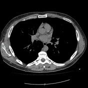
Image CT (lung window) ( update )

Image 3 CT (lung window) ( update )
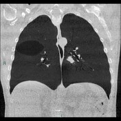
Image 5 CT (lung window) ( update )
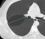
Image 40 CT (lung window) ( destroy )
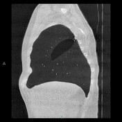
Image 40 CT (lung window) ( create )
Image 48 CT (lung window) ( update )
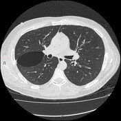
Image 48 CT (lung window) ( destroy )
Updates to Case Attributes
Spontaneousinterlobar pneumothorax is uncommon but early diagnosis can help prevent complications and improve outcomes. Chest X-ray is often diagnostic for a larger pneumothorax, but if the amount of air in the pleural space is minimal, a dedicated CT scan may be required for identification and to resolve diagnostic doubts on the chest radiological examination.
Case courtesy: Dr. Fabio Denicolò, Dr.ssa Eleonora Renzi
Radiographer:TSRM Fabio Imola
-<p>Spontaneous<a href="/articles/interlobar-pneumothorax" title=" interlobar pneumothorax "> interlobar pneumothorax </a>is uncommon but early diagnosis can help prevent complications and improve outcomes. Chest X-ray is often diagnostic for a larger pneumothorax, but if the amount of air in the pleural space is minimal, a dedicated CT scan may be required for identification and to resolve diagnostic doubts on the chest radiological examination.</p><p><br>Case courtesy: Dr. Fabio Denicolò, Dr.ssa Eleonora Renzi</p><p>Radiographer: TSRM Fabio Imola</p>- +<p>Spontaneous <a href="/articles/interlobar-pneumothorax" title=" interlobar pneumothorax ">interlobar pneumothorax</a> is uncommon but early diagnosis can help prevent complications and improve outcomes. Chest X-ray is often diagnostic for a larger pneumothorax, but if the amount of air in the pleural space is minimal, a dedicated CT scan may be required for identification and to resolve diagnostic doubts on the chest radiological examination.</p><p><br>Case courtesy: Dr. Fabio Denicolò, Dr.ssa Eleonora Renzi</p><p>Radiographer: TSRM Fabio Imola</p>


 Unable to process the form. Check for errors and try again.