
Interlobar loculated pneumothorax
Diagnosis certain
Disclosures
- updated 8 Sep 2023:
Nothing to disclose
Updates to Study Attributes
Findings
was changed:
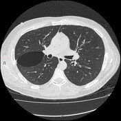
Axial chest CT images show a regularly shaped air loculation, with an obtuse angle between the cystic wall and pleura (length ten cm and anterior-posterior diameter 4 cm). The top and bottom portion of the cystic lesion continues to the right major fissure. Sagittal CT images show the anteroinferior portion of the localised air loculationtapers into the right major fissure. Small apical blebs are also visible on the right adjacent to the aforementioned air loculation. There are initial signs of centrilobular emphysema in the upper lobes. Mediastinum is normal.
Images Changes:
Image 48 CT (lung window) ( create )
Annotation 15825
changed from ,0 arrows,0 labels to blebs ,2 arrows,1 label.


 Unable to process the form. Check for errors and try again.