
Multiple biliary hamartomas
Diagnosis almost certain
Updates to Study Attributes
Findings
was added:
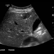
Multiple Biliary Hamartoma. Ultrasound shows many small round echogenic liver lesions and a few larger hypoechoic lesions.
Images Changes:
Image Ultrasound (Transverse) ( update )

Description
was removed:
Single Or Stack Root
was set to
.
Perspective
was set to
Transverse.
Image Ultrasound (Transverse) ( update )
Description
was removed:
Single Or Stack Root
was set to
.
Perspective
was set to
Transverse.
Updates to Study Attributes
Findings
was added:
Multiple Biliary Hamartomas. Portal venous phase CT showing multiple small hypoattenuating liver lesions.
Images Changes:
Image CT (C+ portal venous phase) ( update )

Description
was removed:
Single Or Stack Root
was set to
.
Perspective
was set to
Coronal.
Updates to Study Attributes
Findings
was added:
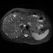
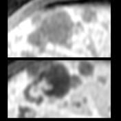
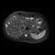
MR images show multiple small T2 hyperintense and T1 hypointense liver lesions. Several of the lesions have an enhancing periphery or enhancing nodule.
Images Changes:
Image MRI (T2 fat sat) ( update )
Description
was removed:
Single Or Stack Root
was set to
.
Image MRI (T1 C+ fat sat) ( update )

Description
was removed:
Single Or Stack Root
was set to
.
Image MRI (T1 C+ fat sat) ( update )
Description
was removed:
Single Or Stack Root
was set to
.
Image MRI (T2 fat sat) ( update )
Description
was removed:
Updates to Case Attributes
Presentation
was changed:
Asymptomatic 35 year old woman.
Age
was set to
35.
Gender
was set to
Female.
Diagnostic Certainty
was set to
.


 Unable to process the form. Check for errors and try again.