
Orbital rhabdomyosarcoma
Diagnosis possible
Disclosures
- updated 11 May 2022:
Nothing to disclose
Updates to Study Attributes
Findings
was changed:
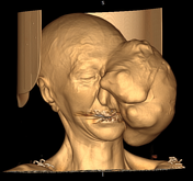
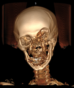
There's a huge mass with necrotic and calcified lesions inside, expanding left orbit, with the displacement of left oculus bulbus to anterioreye globe anteriorly. The mass is involving intothe paranasal sinuses and the nasal cavity. Destruction is seen in the frontal and maxillary bonebones.
Images Changes:
Image CT (VRT) ( update )
Cropped
image
Image CT (VRT) ( update )
Cropped
image
Image CT (non-contrast) ( update )

Cropped
image
Updates to Case Attributes
Body
was changed:
The most probable diagnosis is rhabdomyosarcoma. Neurofibroma is in the differential diagnosis.
-<p>The most probable diagnosis is rhabdomyosarcoma. Neurofibroma is in the differential diagnosis. </p>- +<p>The most probable diagnosis is <a href="/articles/rhabdomyosarcoma-orbit" title="Rhabdomyosarcoma (orbit)">rhabdomyosarcoma</a>. Neurofibroma is in the differential diagnosis. </p>
Systems changed:
- Oncology


 Unable to process the form. Check for errors and try again.