
Osteochondral fracture of the talar dome
Diagnosis certain
Updates to Study Attributes
Modality
was set to
Diagram.
Findings
was added:
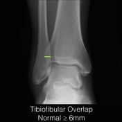
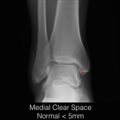
Important normal measurements at the ankle joint.
Tibiofibular overlap should be at least 6mm on an AP image and usually at least 1mm on a mortise view. Reduced overlap is a sign of syndesmotic widening/injury.
Tibiofibular clear space should be less than 6mm on both AP and mortise views. An increase in this space is a sign of syndesmotic widening/injury.
Medial clear space should less than 5mm on both AP and mortise views. An increase in this space is a sign of deltoid ligament injury (assuming there is not a medial malleolus fracture).
Images Changes:
Image 1 Diagram ( create )
Position
was set to
.
Image 2 Diagram ( create )

Image 3 Diagram ( create )


 Unable to process the form. Check for errors and try again.