

Pleural actinomycosis
Updates to Study Attributes
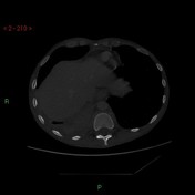
Consolidation within the right lower lobe with loculated right sided pleural effusion.
Multiple enhancing pleural based-based lesions which infiltrate the adjacent fat and involve the right hemidiaphragm.
Abnormal paravertebral soft tissue mass T11-L2 with moth eaten-eaten appearance of T12 vertebral body and associated T12 fracture; abnormal soft tissue narrows the spinal canal at T12.
Appearances are concerning for malignancy. MRI spine and biopsy of pleura advised.
Image CT (bone window) ( update )
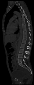
Image 1 CT (lung window) ( update )

Image 3 CT (bone window) ( update )
Updates to Study Attributes
Extensive infiltration of T12 with associated vertebral body collapse.
Pathological soft tissue extends posteriorly to narrow the spinal canal and indents but does not compress the cord.
The infiltrated vertebrae isare surrounded by abnormal soft tissue which iscontiguous with the right pleural mass, suggesting all related to the same disease process.
Small cystic/necrotic regions in the paraspinal soft tissues without drainable abscess.
Summary:Diffuse infiltrating process extending from right basal pleural space to involve T12. Differential involves atypical infection and malignancy.
Updates to Freetext Attributes
Pathology / Microbiology Investigations
The patient underwent ultrasound assisted biopsy of the right pleural mass and a chest drain was inserted tointo the right sided pleural collection.
Microscopy:Sections show multiple core biopsies which consist largely of fibrous connective tissue. There is an infiltrate of mixed inflammatory cells which are predominantly plasma cells but with aggregates of neutrophil polymorphs and scattered lymphocytes. In one core biopsy there is a cluster of actinomyces-like organisms. The overall appearances appear inflammatory in nature and raise the possibility of an infective aetiology.Further info: ISH for Kappa and Lambda show the plasma cell population to be polyclonal. Special stains for organisms highlight Gram positive PAS positive bacterial colonies.
Pleural Fluid:CULTURE RESULT:a) Fusobacterium nucleatum - Isolatedb) Actinomyces meyeri - Isolated


 Unable to process the form. Check for errors and try again.