
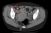
Round ligament leiomyoma
Diagnosis probable
Updates to Study Attributes
Modality
was set to
Annotated image.
Findings
was added:
Pathology highlighted.
Images Changes:
Image Annotated image ( update )
Single Or Stack Root
was set to
.
Image Annotated image ( update )
Single Or Stack Root
was set to
.
Image 1 Annotated image ( update )
Position
was set to
.
Image 2 Annotated image ( update )
Position
was set to
.
Updates to Study Attributes
Modality
was set to
CT.
Findings
was added:
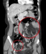
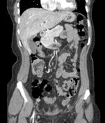
CT images demonstrate a right broad ligament leiomyoma.
Images Changes:
Image CT (C+ portal venous phase) ( update )
Single Or Stack Root
was set to
.
Perspective
was set to
Coronal.
Specifics
was set to
C+ portal venous phase.
Image CT (C+ portal venous phase) ( update )

Single Or Stack Root
was set to
.
Perspective
was set to
Axial.
Specifics
was set to
C+ portal venous phase.
Updates to Case Attributes
Body
was changed:
CT images demonstrate a right broad ligament leiomyoma
This case was donated to Radiopaedia.org by Radswiki.net.
- <p>CT images demonstrate a right broad ligament leiomyoma</p>-- <p>This case was donated to Radiopaedia.org by <a href="/radswikinet-1">Radswiki.net</a></p>- +<p>This case was donated to Radiopaedia.org by <a href="/radswikinet-1">Radswiki.net</a>. </p>
Systems changed:
- Gynaecology


 Unable to process the form. Check for errors and try again.