Serous cystadenoma of the pancreas - macrocystic
Updates to Case Attributes
Distal pancreatectomy and splenectomy
Macroscopic descripton
A distal pancreatectomy specimen comprising pancreas 80x40x20 mm with perpancreatic fat up to 170In an adult female,, and spleen weighing 250 gram. Arising form the posterior pancreatic surface isdifferential for a disrupted unilocular cystic mass 50x35x15 mm fromlesion in the pancreatic margin. The internal lining is smooth. Cyst contents are clearbody and tail includes
-
serous cystadenoma
- typical
- oligocystic/macrocystic
- mucinous cystadenoma
- intraductal papillary mucinous tumour (IPMN)
In this situation, hyalinised fibrous connective tissue. Somethe imaging appearance of the cuboidal cells have clear cytoplasm. Special stain demonstratecystic lesion and the presence of glycogen within the cyst lining cells. No features ofpatient's age is typical for a mucinous epithelial differentiation are recognised (absence of columnar cytology, mucin vacuoles). By immunohistochemistry, the cyst lining cells are positive for epithelial markers CAM 5.2 and AE1/3. There is no staining for neuroendocrine or vascular markers. No ovarian type stroma is identified in H&E sections or immunohistochemistry for oestorgen and progesterone receptors. The peripancreatic lymph nodes have reactive features. The spleen is histologically normal.
Supplementary report
An immunohistochemical stain for CEA reveal no staining in the epithelial liningcystadenoma of the cyst. This support non-mucinous differentiation of the epitheliumpancreas, but on pathologic analysis, an oligocystic/macrocystic cystadenoma was diagnosed.
Diagnosis
Benign pancreatic cyst, favour macrocystic serous cystadenoma
-<p>Distal pancreatectomy and splenectomy</p>-<p><strong>Macroscopic descripton</strong></p>-<p>A distal pancreatectomy specimen comprising pancreas 80x40x20 mm with perpancreatic fat up to 170,, and spleen weighing 250 gram. Arising form the posterior pancreatic surface is a disrupted unilocular cystic mass 50x35x15 mm from the pancreatic margin. The internal lining is smooth. Cyst contents are clear and not mucoid. The cyst does not communicate with pancreatic duct.</p>-<p><strong>Microscopic description</strong></p>-<p>Sections show a unilocular cyst surrounded by normal pancreatic parenchyma. The cyst is lined by a single layer of bland low cuboidal cells supported by dense, hyalinised fibrous connective tissue. Some of the cuboidal cells have clear cytoplasm. Special stain demonstrate the presence of glycogen within the cyst lining cells. No features of mucinous epithelial differentiation are recognised (absence of columnar cytology, mucin vacuoles). By immunohistochemistry, the cyst lining cells are positive for epithelial markers CAM 5.2 and AE1/3. There is no staining for neuroendocrine or vascular markers. No ovarian type stroma is identified in H&E sections or immunohistochemistry for oestorgen and progesterone receptors. The peripancreatic lymph nodes have reactive features. The spleen is histologically normal.</p>-<p><strong>Supplementary report</strong></p>-<p>An immunohistochemical stain for CEA reveal no staining in the epithelial lining of the cyst. This support non-mucinous differentiation of the epithelium.</p>-<p><strong>Diagnosis</strong></p>-<p>Benign pancreatic cyst, favour macrocystic serous cystadenoma</p>- +<p>In an adult female, the differential for a cystic lesion in the pancreatic body and tail includes</p><ul>
- +<li>
- +<a title="Serous cystadenoma of the pancreas" href="/articles/serous-cystadenoma-of-pancreas">serous cystadenoma</a><ul>
- +<li>typical</li>
- +<li>oligocystic/macrocystic</li>
- +</ul>
- +</li>
- +<li><a title="Mucinous cystadenoma of the pancreas" href="/articles/mucinous-cystadenoma-of-pancreas">mucinous cystadenoma</a></li>
- +<li><a title="IPMN" href="/articles/intraductal-papillary-mucinous-neoplasm">intraductal papillary mucinous tumour (IPMN)</a></li>
- +</ul><p>In this situation, the imaging appearance of the cystic lesion and the patient's age is typical for a mucinous cystadenoma of the pancreas, but on pathologic analysis, an oligocystic/macrocystic cystadenoma was diagnosed.</p>
Tags changed:
- pancreas
Updates to Freetext Attributes
Distal pancreatectomy and splenectomy
Macroscopic descripton
A distal pancreatectomy specimen comprising pancreas 80x40x20 mm with perpancreatic fat up to 170,, and spleen weighing 250 gram. Arising form the posterior pancreatic surface is a disrupted unilocular cystic mass 50x35x15 mm from the pancreatic margin. The internal lining is smooth. Cyst contents are clear and not mucoid. The cyst does not communicate with pancreatic duct.
Microscopic description
Sections show a unilocular cyst surrounded by normal pancreatic parenchyma. The cyst is lined by a single layer of bland low cuboidal cells supported by dense, hyalinised fibrous connective tissue. Some of the cuboidal cells have clear cytoplasm. Special stain demonstrate the presence of glycogen within the cyst lining cells. No features of mucinous epithelial differentiation are recognised (absence of columnar cytology, mucin vacuoles). By immunohistochemistry, the cyst lining cells are positive for epithelial markers CAM 5.2 and AE1/3. There is no staining for neuroendocrine or vascular markers. No ovarian type stroma is identified in H&E sections or immunohistochemistry for oestorgen and progesterone receptors. The peripancreatic lymph nodes have reactive features. The spleen is histologically normal.
Supplementary report
An immunohistochemical stain for CEA reveal no staining in the epithelial lining of the cyst. This support non-mucinous differentiation of the epithelium.
Diagnosis
Benign pancreatic cyst, favour macrocystic serous cystadenoma
Updates to Study Attributes
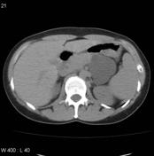
There is a large (~5 x 5cm5 cm) well circumscribed-circumscribed, homogeneous lesion with a clear margin and fluid attenuation in the tail of the pancreas adjacentadjacent to the spleen and left kidney. It has a clear margin and shows fluid attenuation, suggestive of a cystic lesion. The pancreas is otherwise unremarkable.
Following administration of contrast there is no convincing solid component to the mass. No calcifications.
Image CT (C+ arterial phase) ( update )

Image CT (non-contrast) ( update )


 Unable to process the form. Check for errors and try again.