
Traumatic neuroma - ulnar nerve at elbow
Diagnosis almost certain
Disclosures
- updated 2 Aug 2023:
Nothing to disclose
Updates to Study Attributes
Caption
was added:
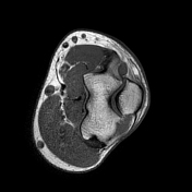
Left elbow MRI with contrast
Modality
changed from to MRI.
Findings
was added:
A defect within the cubital tunnel retinaculum measuring 5 mm in length (Blue line, image A) with evidence of ulnar nerve fascicles outpouching through the defect measures 14x4 mm (Yellow outline, image A).
Just distally there is partial nerve injury with loss of the medial portion nerve fascicles at the level of cubital tunnel (Red arrow, image B) followed by focal nodular circumferential thickening of the ulnar nerve just distal to the cubital tunnel measures 7x8x11 mm with circumferential of measures 33 mm2 (in comparison with the normal circumferential diameter of about 8 mm2) denoting central traumatic neuroma (White arrow, image C).
Post contrast images show no abnormal enhancement.
Images Changes:
Image 1 MRI (PD fat sat) ( update )

Position
changed from 4 to 1.
Image 2 MRI (T1) ( update )
Position
changed from 4 to 2.
Image 7 MRI (T2 Dixon) ( update )

Position
changed from 5 to 7.


 Unable to process the form. Check for errors and try again.