


121,135 results
Case
Marginal cord insertion

Published
29 Sep 2023
97% complete
Ultrasound
Case
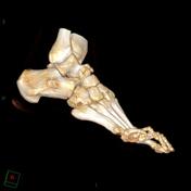
Os supranaviculare
Published
31 Oct 2011
71% complete
CT
Case
Mesothelioma - subtle nodular thickening of the interlobar fissures

Published
09 May 2018
92% complete
CT
Annotated image
X-ray
Case
Finger PIP joint septic arthritis and osteomyelitis

Published
01 Sep 2020
94% complete
X-ray
Article
Mid-talar axis
The mid-talar axis represents a line drawn down the longitudinal axis of the talus and can be drawn on lateral and DP radiographs.
Measurement
Independent on the view on which the line is drawn, it should bisect the neck of the talus and the head.
On the lateral and DP views, the line should...
Case
Achilles tendon injury

Published
02 May 2014
66% complete
X-ray
Ultrasound
Case
Tongue stud

Published
01 Jan 2009
59% complete
CT
Case
PASTA Lesion

Published
14 Mar 2018
89% complete
MRI
Case
Gastric carcinoma

Published
27 Jul 2014
71% complete
CT
Case
Stieda fracture

Published
19 Jan 2022
91% complete
X-ray
Case
Steinstrausse - multilevel

Published
20 May 2016
92% complete
CT
Case
Bone infarct of humerus

Published
20 Aug 2016
75% complete
X-ray
Article
Perivascular spaces
Perivascular spaces, also known as Virchow-Robin spaces, are fluid-filled spaces that surround small arterioles, capillaries and venules in the brain. Those that surround perforating vessels are frequently seen on routine MRI imaging.
Despite having been described well over a century ago and se...
Case
Diffuse large B-cell lymphoma (DLBCL)

Published
15 Aug 2023
74% complete
MRI
CT
Annotated image
Case
Pneumomediastinum and subcutaneous emphysema

Published
05 May 2013
91% complete
X-ray
Case
Peripelvic cysts

Published
09 Jul 2022
76% complete
CT
Article
Parastomal hernia
Parastomal hernias (alternative plural: herniae) are defined as the protrusion of abdominal contents through an abdominal wall defect in the vicinity of the stoma.
Classification
The hernia may contain a loop of bowel forming the stoma itself, omentum, and/or intestinal loops other than that f...
Article
MitraClip device
A MitraClip is a device for percutaneous mitral valve repair. It is a percutaneous edge-to-edge attachment system that mimics the surgical procedure. This technique creates a tissue bridge between the anterior and posterior leaflets, employing one clip deployed through transseptal catheterisatio...
Case
Acromioclavicular joint ganglion and long head of biceps brachii dislocation

Published
06 Feb 2011
63% complete
Ultrasound
Article
Benign and malignant characteristics of breast lesions at ultrasound
Benign and malignant characteristics of breast lesions at ultrasound allow the classification as either malignant, intermediate or benign based on work published by Stavros et al. in 1995.
Radiographic features
Ultrasound
Malignant characteristics (with positive predictive values)
sonographi...


 Unable to process the form. Check for errors and try again.