


4,572 results found
Case
Rectus femoris muscle injury with "Bull's eye" sign

Published
28 Feb 2023
77% complete
MRI
Case
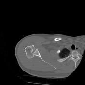
Posterior shoulder dislocation
Published
27 Sep 2021
86% complete
CT
Article
Tension gastrothorax
Tension gastrothorax describes a rare life-threatening condition caused by mediastinal shift due to a distended stomach herniating into the thorax through a diaphragmatic defect.
Clinical presentation
Presentation is generally with acute and severe respiratory failure, with clinical features ...
Case
Lisfranc injury

Published
18 Aug 2017
89% complete
CT
Case
Lover's fracture

Published
12 Feb 2014
82% complete
X-ray
Article
Craniocervical fixation
Craniocervical fixation, instrumentation or occipitocervical fusion refer to surgical fixation techniques with the goal to stabilise the craniocervical junction.
Indications
Craniocervical fixation is indicated in the setting of craniocervical instability including 2,3:
iatrogenic craniocervi...
Article
Spinal compression fracture
Spinal compression fractures occur as a result of injury, commonly fall onto the buttock or pressure from normal activities, to the weakened vertebrae due to osteoporosis.
Epidemiology
They have a reported incidence of 1.2 per 1000 person-years after 85 years of age in the United States. Howev...
Case
Absent distal humeral shaft post trauma

Published
06 Sep 2018
94% complete
X-ray
Article
Metal foreign body
Metal foreign bodies may be present if they are ingested, inserted, or as a result of an injury.
Radiographic features
Nearly all metals are radiopaque and can be seen on plain radiographs and CT with the exception of aluminium, which may not be seen on plain radiographs 1,2.
Ultrasound
Met...
Case
Chronic penetrating traumatic brain injury - intracranial bullet

Published
12 Mar 2020
73% complete
CT
Article
Humeral shaft fracture
Humeral shaft fractures are readily diagnosed and usually, do not require internal fixation.
Epidemiology
Humeral shaft fractures account for 3-5% of all fractures 1,3. Although they occur in all age groups, a bimodal distribution is noted. The first peak is seen in the third decade in males ...
Case
Thyroid cartilage fracture

Published
27 Feb 2015
74% complete
CT
Case
Pancreatic transection

Published
12 Mar 2014
90% complete
Annotated image
CT
Case
Luxatio erecta and greater tubercle fracture

Published
04 Dec 2019
91% complete
X-ray
Article
Anderson and Montesano classification of occipital condyle fractures
The Anderson and Montesano classification is a widely used system for describing occipital condyle fractures. It divides injuries into three types based on morphology and mechanism of injury 1-5.
Classification
type I: impacted type occipital condyle fracture
morphology: comminution of the co...
Case
Bony bankart and Hill-Sach defect

Published
29 Nov 2021
95% complete
CT
MRI
Case
Occipital condyle fracture (type 1) and atlas transverse process fracture (type 5)

Published
17 Mar 2021
92% complete
CT
Case
Subcapital femoral neck fracture

Published
22 Nov 2012
79% complete
X-ray
Article
Avulsion injury
Avulsion injuries or fractures occur where the joint capsule, ligament, tendon or muscle attachment site is pulled off from the bone, usually taking a fragment of cortical bone. Avulsion fractures are commonly distracted due to the high tensile forces involved. There are numerous sites at which ...
Case
Scalp haematoma types (diagram)

Published
30 Apr 2018
38% complete
Diagram


 Unable to process the form. Check for errors and try again.