


2,038 results
Article
Ligamentum teres hepatis (abdomen)
The ligamentum teres hepatis or round ligament is the fibrous cord formed by the obliterated fetal umbilical vein that runs in the free edge of the falciform ligament from the umbilicus into the left lobe of the liver.
Article
CT abdomen-pelvis (protocol)
The CT abdomen-pelvis protocol serves as an outline for an examination of the whole abdomen including the pelvis. It is one of the most common CT protocols for any clinical questions related to the abdomen and/or in routine and emergencies. It forms also an integral part of trauma and oncologic ...
Article
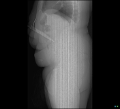
Paediatric abdomen (PA erect view)
The PA erect abdominal radiograph is the standard view for assessing air-fluid levels and free air in the paediatric abdomen. This view may be taken alongside the AP supine and lateral decubitus views. As radiation protection is an essential consideration in paediatrics, some departmental protoc...
Article
Paediatric abdomen (lateral decubitus view)
The lateral decubitus radiograph is an additional projection for assessing the paediatric abdomen. This view is ideal for displaying free gas in the abdomen and/or if the patient is unable to lie supine 1. As radiation dose is an important consideration for paediatric imaging, the lateral decubi...
Article
Paediatric abdomen (AP supine view)
The AP supine abdominal radiograph is a routine view when imaging the paediatric abdomen. This view may be taken alongside the PA erect and lateral decubitus views. As radiation protection is an essential consideration in paediatrics, some departmental protocols may only perform one view (either...
Article
CT chest abdomen-pelvis (protocol)
The CT chest-abdomen-pelvis protocol serves as an outline for an examination of the trunk covering the chest, abdomen and pelvis. It is one of the most common CT examinations conducted in routine and emergencies. It can be combined with a CT angiogram.
Note: This article aims to frame a genera...
Article
CT neck, chest, abdomen-pelvis (NCAP protocol)
The CT neck chest-abdomen-pelvis protocol aims to evaluate the neck, thoracic and abdominal structures using contrast in trauma imaging. The use of contrast facilitates the assessment of pathologies globally whilst minimising dose by potentially disregarding a non-contrast scan.
Note: This art...
Article
Paediatric abdomen (prone cross-table lateral view)
The prone cross-table lateral view is an additional projection to demonstrate the paediatric abdomen and is a more ideal alternative to the invertogram, which may be less comfortable for the patient. This discomfort may result in a continuously crying baby, causing the puborectalis sling to cont...
Article
Paediatric abdomen (supine cross-table lateral view)
The supine cross-table lateral view is an additional projection to demonstrate the paediatric abdomen. As radiation dose is an important consideration for paediatric imaging, the horizontal beam lateral view is not often performed; although this will vary based on the department.
Indications
T...
Case
Gunshot injury to the abdomen

Published
09 Feb 2018
98% complete
CT
Case
Normal CTA abdomen and pelvis
Published
21 Feb 2024
95% complete
CT
Case
Normal CT abdomen and pelvis - female

Published
21 Jul 2023
95% complete
CT
Case
Tuberculous lymphadenitis - abdomen

Published
18 Apr 2020
80% complete
Ultrasound
CT
Case
Chest and abdomen multi-trauma

Published
11 Dec 2013
92% complete
CT
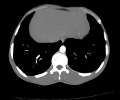
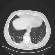
Case
COVID-19 pneumonia (CT abdomen)
Published
26 Mar 2020
89% complete
CT
Case
Sarcoidosis in the abdomen and pelvis

Published
16 Apr 2019
74% complete
CT
Case
Metallic foreign body in abdomen

Published
25 Apr 2014
79% complete
X-ray
Case
Knife penetrating abdomen indenting the IVC
Published
08 Oct 2023
85% complete
CT
Case
Normal MRI abdomen in pregnancy

Published
23 Mar 2021
77% complete
MRI
Case
Complicated cholecystitis with gallbladder perforation and diffuse acute abdomen

Published
15 Jul 2014
90% complete
Annotated image
CT


 Unable to process the form. Check for errors and try again.