


5,943 results
Article
Multiple endocrine neoplasia type IIb
Multiple endocrine neoplasia (MEN) type IIb, also known as MEN type 3 (MEN3) 3 or mucosal neuroma syndrome 2, accounts for only 5% cases of MEN2 and is characterised by:
phaeochromocytoma(s): in 50% of patients, often bilateral, and can be extra-adrenal
medullary thyroid cancer: 100% of patien...
Article
Crossed renal ectopia
Crossed renal ectopia is said to be present when the kidney is seen in the opposite retroperitoneal space. It is more common for the left kidney to be ectopically located on the right side. More than 85% of these get fused resulting in crossed fused renal ectopia. Less than 15% cases are non-fus...
Case
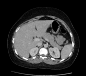
Renal artery aneurysm causing focal hydronephrosis

Published
13 Feb 2024
95% complete
CT
Case
5-alpha reductase deficiency
Published
17 Jun 2024
59% complete
CT
Case
Renal cell carcinoma with dominant necrotic changes

Published
14 Mar 2024
77% complete
CT
Case
Partial segmental thrombosis of the corpus cavernosum

Published
24 Jun 2024
74% complete
MRI
Article
Pubococcygeal line
The pubococcygeal line (PCL) is a reference line for the pelvic floor on imaging studies and helps detect and grade pelvic floor prolapse in defaecography studies.
Measurement
The pubococcygeal line is defined as one that connects the inferior border of the symphysis pubis (anterior margin) t...
Case
Testicular seminoma

Published
26 Jan 2024
94% complete
Ultrasound
Case
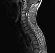
Von Hippel-Lindau disease with spinal haemangioblastoma
Published
17 Sep 2010
71% complete
MRI
Ultrasound
Case
Seminal vesicle cyst

Published
29 Apr 2024
92% complete
MRI
Case
Gossypiboma

Published
17 Feb 2024
95% complete
CT
Article
TWIST score
The Testicular Workup for Ischaemia and Suspected Torsion (TWIST) score is a clinical decision tool used for the workup and management of acute scrotal emergencies where testicular torsion is suspected. It uses history and examination to estimate the likelihood of torsion. Validation of the clin...
Case
Whirlpool (illustration)

Published
12 Feb 2024
35% complete
Photo
Article
Diverticulum
Diverticula are outpouchings of a hollow viscus and can be either true or false.
Occasionally a diverticulum is used in a more general sense to mean the outpouching of other anatomical structures, e.g. frontal intersinus septal cells are hypothesised to form as diverticula from the frontal sinu...
Case
Urethral diverticulum
Published
23 Jun 2024
94% complete
Fluoroscopy
Case
Medullary nephrocalcinosis

Published
13 Mar 2017
89% complete
Ultrasound
X-ray
CT
Case
Lower anterior abdominal wall pericatheter collection (CT peritoneography)

Published
05 Sep 2023
87% complete
CT
Case
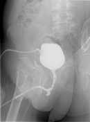
Manta ray sign - bladder exstrophy

Published
15 Mar 2024
94% complete
Fluoroscopy
Article
Manta ray sign (bladder)
The manta ray sign is a radiographic appearance in bladder exstrophy. It describes wide midline separation of the pubic bones simulating the appearance of a manta ray swimming towards you 1. The sacrum and iliac wings recall the manta ray’s head and body, with the widely spaced pubic rami formin...
Case
Intravesical ureterocele

Published
27 Jun 2024
74% complete
CT


 Unable to process the form. Check for errors and try again.