


3,794 results
Case
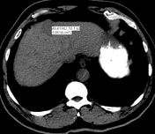
Absent portal vein bifurcation

Published
13 Jan 2020
89% complete
CT
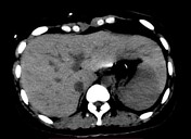
Case
Beaver tail liver
Published
03 Jan 2023
73% complete
CT
Article
Gallbladder folds
Gallbladder folds arise due to the gallbladder wall folding onto itself. They are thick, junctional in nature and incomplete or non-continuous in appearance.
The posterior wall is usually involved, however, anterior wall folds may also occur 1. The folding may produce a bizarre or unusual shap...
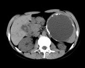
Case
Biliary cystadenoma

Published
20 Oct 2010
74% complete
CT
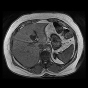
Case
Focal hepatic steatosis
Published
24 Jan 2022
89% complete
MRI
Case
Hepatocellular carcinoma

Published
06 Dec 2020
78% complete
CT
MRI
Article
Pancreatic calcifications
Pancreatic calcifications can arise from many aetiologies.
Punctate intraductal calcifications
chronic pancreatitis
alcoholic pancreatitis (20-40%) 2
intraductal, numerous, small, irregular
preponderant cause of diffuse pancreatic intraductal calcification
gallstone pancreatitis (2%) 2
...
Article
Hepatic haemangiomatosis
Hepatic haemangiomatosis is a condition in which there are multiple haemangiomas affecting the liver.
Terminology
When the lesions are spread throughout the liver, then this is termed diffuse hepatic haemangiomatosis.
Pathology
Associations
giant liver haemangioma 2
Radiographic features
...
Article
Ectopic pancreatic tissue
Ectopic pancreatic tissue, also known as heterotopic pancreatic tissue, refers to the presence of pancreatic tissue in the submucosal, muscularis or subserosal layers of the luminal gastrointestinal tract outside the normal confines of the pancreas and lacking any anatomic or vascular connection...
Case
Gallbladder carcinoma
Published
06 Jan 2019
77% complete
CT
Case
Hepatic hydatid cyst - CE 3B

Published
29 Apr 2024
80% complete
CT
Case
Breast cancer pseudocirrhosis with lobar invovlement

Published
02 Sep 2020
80% complete
CT
Case
Hydatid cyst of the spleen
Published
28 Aug 2010
62% complete
CT
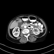
Case
Polysplenia syndrome

Published
24 May 2021
86% complete
CT
Article
Hepatocystic triangle
The hepatocystic triangle (or Calot triangle) is a small triangular space at the porta hepatis of surgical importance as it is dissected during cholecystectomy. Its contents, the cystic artery and cystic duct, must be identified before ligation and division to avoid intraoperative injury.
Gros...
Case
Hepatocellular carcinoma

Published
29 Oct 2023
80% complete
MRI
Case
Gallbladder polyp

Published
15 May 2017
82% complete
Ultrasound
Diagram
Case
Cirrhosis

Published
21 May 2021
95% complete
CT
Case
Ciliated hepatic foregut duplication cyst
Published
02 Dec 2017
50% complete
CT
Article
Klebsiella
Klebsiella is a genus of Gram-negative, oxidase-negative, rod-shaped bacteria, which is relatively commonly encountered in the healthcare environment. It has numerous species, including K. pneumoniae, K. aerogenes, and K. rhinoscleromatis 1. Klebsiella may cause a range of infections, most commo...


 Unable to process the form. Check for errors and try again.