


5,952 results
Article
Vesicourachal diverticulum
Vesicourachal diverticulum, or just urachal diverticulum, is one of the congenital urachal remnant abnormalities.
Gross anatomy
It is the proximal equivalent of a urachal umbilical sinus, representing a result of the failure of the urachus to close at the urinary bladder, forming an out-pouchi...
Article
Urachal cyst
Urachal cysts are one of the manifestations of the spectrum of congenital urachal remnant abnormalities.
Epidemiology
An infected urachal cyst can occur at any age.
Clinical presentation
Urachal cysts usually remain asymptomatic until complicated by infection or bleeding.
Pathology
Uracha...
Case
Complications of sickle cell disease

Published
25 Sep 2023
93% complete
CT
X-ray
Fluoroscopy
Case
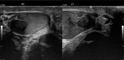
Epididymal cyst

Published
27 Mar 2023
72% complete
Ultrasound
Case
Metastatic endometrial cancer
Published
10 Feb 2024
81% complete
CT
MRI
Article
Ureteral pseudodiverticulosis
Ureteral pseudodiverticulosis are acquired false diverticula resulting from herniation of epithelium through the muscularis layer of the ureter and characterized by the presence of multiple outpouchings smaller than 5 mm. It is sometimes bilateral and is often located in the upper two-thirds of ...
Case
Renal hemorrhage
Published
10 Feb 2024
95% complete
DSA (angiography)
CT
Case
Urothelial cell carcinoma of renal collecting system

Published
05 Feb 2024
74% complete
CT
Case
Hot kidneys (bone scan)

Published
19 May 2024
66% complete
Nuclear medicine
Case
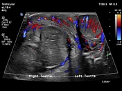
Testicular torsion
Published
10 Feb 2024
94% complete
Ultrasound
Case
Wilms tumor

Published
19 May 2024
72% complete
Ultrasound
Case
Bifid collecting system with ureterocele

Published
13 Mar 2024
94% complete
Fluoroscopy
Article
Cobra head sign (ureter)
The cobra head sign, also known as the spring onion sign, refers to dilatation of the distal ureter, surrounded by a thin lucent line seen in patients with an adult-type ureterocele. The cobra head appearance indicates an uncomplicated ureterocele.
During an excretory phase of an intravenous ur...
Case
Prostatic abscesses

Published
23 Jan 2024
80% complete
MRI
Case
Urachal adenocarcinoma

Published
04 Feb 2024
77% complete
CT
Case
Nutcracker syndrome with varicocele
Published
26 Feb 2024
75% complete
Ultrasound
Case
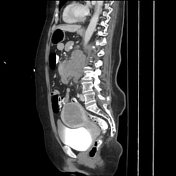
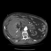
Spontaneous rupture of renal cell carcinoma

Published
13 Dec 2020
92% complete
CT
Ultrasound
Case
Testicular adrenal rests
Published
10 Feb 2024
79% complete
Ultrasound
Case
Renal laceration (collecting system involvement)

Published
21 Mar 2013
92% complete
CT
Article
Lobar nephronia
Lobar nephronia, also known as acute focal nephritis, refers to an intermediate stage between acute pyelonephritis and renal abscess, and is a focal region of interstitial nephritis.
It appears as a wedge of poorly perfused renal parenchyma, without a cortical rim sign.
The condition is discu...


 Unable to process the form. Check for errors and try again.