


121,251 results
Article
Gallbladder perforation
Gallbladder perforations are a serious complication of acute cholecystitis and represent an advanced stage of the disease. They tend to occur in an elderly and/or comorbid demographic and carry higher rates of morbidity and mortality.
Clinical presentation
Symptoms and clinical signs are varia...
Article
Inverted papilloma
Inverted papillomas are a type of Schneiderian papilloma, representing an uncommon non-cancerous sinonasal tumor that mostly affects middle-aged men. They may rarely undergo malignant transformation, most commonly into squamous cell carcinoma. On imaging, they classically demonstrate a convolute...
Case
Multiple liver adenomas with signs of bleeding

Published
04 Dec 2014
87% complete
CT
MRI
Article
Psoriatic arthritis
Psoriatic arthritis (PsA) is inflammatory arthritis associated with psoriasis. It is usually negative for rheumatoid factor and hence classified as one of the seronegative spondyloarthritides.
Epidemiology
Overall prevalence is ~0.5% (range 0.1-1%), however, it affects up to ~25% (range 6-41%)...
Article
Capsulolabral insertion classification
Capsulolabral insertion classification reflects the variation in the relationship between the glenoid labrum and the attachment of the anteroinferior shoulder joint capsule.
Classification
This classification was originally described by Mosely and Övergaard in 1962 on cadavers 3 but has since ...
Case
Ectopic posterior pituitary

Published
12 Feb 2022
80% complete
MRI
Article
Black hole sign (intracerebral hemorrhage)
The black hole sign refers to the non-contrast CT appearance of acute extravasation of blood into a hematoma, for example, an intracerebral hemorrhage, and therefore is a predictor of hemorrhage expansion 3. It can be thought of as an encapsulated swirl sign.
Radiographic features
The black ho...
Case
Acute mesenteric ischemia

Published
12 Feb 2022
89% complete
CT
Case
Posteromedial corner injury

Published
25 Dec 2009
62% complete
CT
X-ray
MRI
Case
Cleft palate

Published
20 Jun 2013
63% complete
X-ray
Case
Proctosigmoiditis

Published
09 May 2020
92% complete
CT
Diagram
Article
Tracheal air column
The tracheal air column describes the appearance of the trachea on plain chest radiographs.
Radiographic appearance
Plain radiograph
On frontal chest radiographs, the air column extends as an almost vertical, radiolucent column midline in the mediastinum from the inferior margin of the cricoi...
Case
Lumbar disc extrusion

Published
13 Sep 2008
56% complete
Annotated image
CT
Case
Fat-water swap artifact

Published
06 Mar 2020
50% complete
MRI
Article
Ossifying fibroma
Ossifying fibromas are benign bone lesions that should be differentiated from non-ossifying fibromas and fibrous dysplasia. Osteofibrous dysplasia is considered as a separate pathological entity in view of its different presentation and treatment, although histopathologically similar to ossifyin...
Case
Focal fatty sparing of the liver

Published
07 Sep 2019
74% complete
MRI
Case
Scleroderma
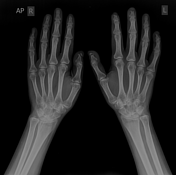
Published
09 Sep 2018
97% complete
X-ray
Case
Rheumatoid arthritis - elbow

Published
06 Mar 2024
92% complete
MRI
Case
Cystolithiasis

Published
26 May 2023
82% complete
Ultrasound
X-ray
Case
Complete ACL and MCL tears with lateral meniscus transection

Published
09 Jan 2021
77% complete
MRI


 Unable to process the form. Check for errors and try again.