
Cases
By sharing our collective experience through interesting and classic patient cases, we can make a real difference in how people are imaged and diagnosed. Each case belongs to a contributing member and all cases are reviewed by our dedicated editors to ensure they reach quality standards and abide by privacy guidelines. Cases can public or unlisted and then be viewed directly or added to articles, playlists or multiple choice questions. Find out more about cases.


1,701 results found
Case
Wernicke encephalopathy

Published
16 Feb 2022
80% complete
MRI
Diagram
Case
Chondrocalcinosis: diagram of causes

Published
01 Oct 2012
32% complete
Diagram
Case
True bovine arch (illustration)

Published
10 May 2015
22% complete
Diagram
Case
Platybasia (diagrams)

Published
07 May 2008
32% complete
Diagram
Case
Benign enlargement of the subarachnoid spaces (BESS)

Published
09 Jan 2020
98% complete
Diagram
MRI
Case
Mini-gastric bypass surgery
Published
07 Sep 2020
62% complete
Diagram
CT
Case
Geometric blur (illustrations)

Published
14 Mar 2024
41% complete
Diagram
Case
Foreign bodies (finger)

Published
16 Mar 2024
85% complete
Ultrasound
Diagram
Photo
Case
Decision tree

Published
25 Apr 2019
44% complete
Diagram
Case
Spinal cord (Gray's illustration)

Published
07 Sep 2020
35% complete
Diagram
Case
Lymph node

Published
09 Dec 2020
19% complete
Diagram
Case
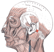
Posterior auricular muscle (Gray's illustration)

Published
27 Jun 2023
29% complete
Diagram
Case
Posterior auricular muscle (Gray's illustration)
Published
27 Jun 2023
29% complete
Diagram
Case
Inguinal canal and rings (Gray's illustrations)

Published
03 Aug 2021
32% complete
Diagram
Case
Slipped upper femoral epiphysis

Published
02 Feb 2010
88% complete
X-ray
Diagram
Case
Brainstem motor nuclei - Gray's anatomy illustration

Published
17 May 2015
35% complete
Diagram
Case
Midbrain (axial) showing tectum and tegmentum

Published
16 May 2015
38% complete
Diagram
Case
Subarachnoid cistern (illustration)

Published
16 May 2015
25% complete
Diagram
Case
Cerebellum (sagittal) - Gray's anatomy illustration

Published
16 May 2015
38% complete
Diagram
Case
Biceps femoris muscle long head (Gray's illustration)

Published
16 Dec 2022
44% complete
Diagram
ADVERTISEMENT: Supporters see fewer/no ads


 Unable to process the form. Check for errors and try again.