
Search results for “abdominal ct ”


73 results found
Question
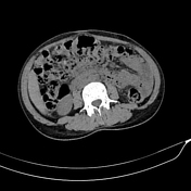
Question 1635
A 10-year-old boy presents in the emergency department with complaints of sudden right lower abdominal pain. CT image is presented. What is the most likely diagnosis?
Article
Endometrioma
Endometriomas, also known as chocolate cysts or endometriotic cysts, are a localized form of endometriosis and are usually within the ovary. They are readily diagnosed on ultrasound, with most demonstrating classical radiographic features.
Epidemiology
These occur in up to 10% of women of rep...
Article
CT abdomen-pelvis (protocol)
The CT abdomen-pelvis protocol serves as an outline for an examination of the whole abdomen including the pelvis. It is one of the most common CT protocols for any clinical questions related to the abdomen and/or in routine and emergencies. It forms also an integral part of trauma and oncologic ...
Article
Acute appendicitis
Acute appendicitis is an acute inflammation of the vermiform appendix. It is a very common condition in general radiology practice and is one of the main reasons for abdominal surgery in young patients. CT is the most sensitive modality to detect appendicitis.
Terminology
Acute appendicitis (p...
Article
Cesarian scar endometriosis
Cesarian scar endometriosis can be located in the skin, subcutaneous tissue, rectus muscle/sheath, intraperitoneally, or in the uterine myometrium (within uterine scar).
Epidemiology
The reported incidence of abdominal scar endometriosis following cesarean section is 0.03-0.6% 6.
Clinical pre...
Question
Question 1623
A 45-year-old patient presents with abdominal pain and vomiting. A CT of the abdomen is performed. An arterial-phase image is shown. What is the most likely diagnosis?
Article
Acute abdominal pain
Acute abdominal pain is a common acute presentation in clinical practice. It encompasses a very broad range of possible etiologies and diagnoses, and imaging is routinely employed as the primary investigative tool in its modern management.
Terminology
A subgroup of patients with acute abdomina...
Case
Ovarian mucinous cystadenofibroma

Published
04 Dec 2022
95% complete
CT
Photo
Case
Incidental invasive lobular carcinoma and incidental mature ovarian teratoma

Published
10 May 2014
86% complete
Annotated image
Mammography
CT
Article
Missing IUCD
A missing IUCD is considered when the retrieval strings of certain types of intrauterine contraception devices (IUCD) cannot be seen on physical examination. The possibilities are
expulsion of IUCD
migration of IUCD
detachment of IUCD thread
uterine perforation in IUCD
embedd...
Question
Question 1620
This 50-year-old man presents with abdominal pain. Abdominal x-ray shows small bowel dilatation. Contrast-enhanced CT is performed with selected images shown. What is the most likely diagnosis?
Case
Ovarian torsion

Published
06 Dec 2021
83% complete
CT
Case
Hemorrhagic ovarian follicle cyst (CT)

Published
11 Sep 2022
80% complete
CT
Case
Complete hydatidiform mole with bilateral theca lutein cysts

Published
08 Mar 2015
83% complete
Ultrasound
CT
Case
Mature cystic teratoma of ovary

Published
29 Dec 2012
89% complete
CT
Case
Pelvic septic thrombophlebitis
Published
18 Oct 2023
77% complete
CT
Case
Malignant mixed Mullerian tumor of the uterus

Published
11 May 2020
95% complete
CT
Ultrasound
Article
Acute non-traumatic abdominal pain in pregnancy
Acute non-traumatic abdominal pain in pregnancy requires a considered imaging approach due to the increased risks of fetal demise associated with undiagnosed diseases such as perforated acute appendicitis. Ultrasound is the first-line modality due to its wide availability and ability to diagnose...
Case
Menstrual cup (CT)

Published
17 Feb 2021
59% complete
Photo
CT
Case
Rectus abdominis hematoma following cesarean section

Published
05 Oct 2019
80% complete
Ultrasound
CT


 Unable to process the form. Check for errors and try again.