


201 results found
Case
Sternomanubrial dislocation (diagrams)

Published
27 Dec 2023
44% complete
Diagram
Case
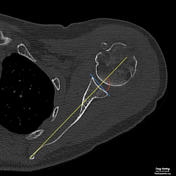
Glenoid version measurement - Friedman method (diagram)
Published
21 Dec 2022
44% complete
Diagram
Case
Sacral plexus (Gray's illustrations)

Published
11 Oct 2022
35% complete
Diagram
Case
Leg and foot nerves (Gray's illustrations)

Published
11 Oct 2022
35% complete
Diagram
Case
Lower limb nerves (Gray's illustrations)

Published
10 Oct 2022
35% complete
Diagram
Case
Lumbar plexus (Gray's illustrations)

Published
06 Oct 2022
32% complete
Diagram
Case
Upper limb nerves (Gray's illustrations)

Published
02 Feb 2022
35% complete
Diagram
Case
Thoracic cutaneous nerves (Gray's illustration)

Published
02 Feb 2022
32% complete
Diagram
Case
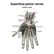
Palmar nerves (Gray's illustrations)
Published
27 Jan 2022
35% complete
Diagram
Case
Intercostal nerves (Gray's illustrations)

Published
27 Jan 2022
32% complete
Diagram
Case
Brachial plexus (Gray's illustrations)

Published
05 Jan 2022
32% complete
Diagram
Case
Cutaneous spinal nerves of the upper limb (Gray's illustrations)

Published
05 Jan 2022
29% complete
Diagram
Case
Origins of the extraocular muscles (Gray's illustration)

Published
27 Dec 2021
32% complete
Diagram
Case
Innervation of the medial and lateral recti muscles (Gray's illustration)

Published
27 Dec 2021
35% complete
Diagram
Case
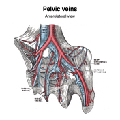
Pelvic veins (Gray's illustration)
Published
20 Dec 2021
32% complete
Diagram
Case
Superficial abdominal wall veins (Gray's illustration)

Published
20 Dec 2021
29% complete
Diagram
Case
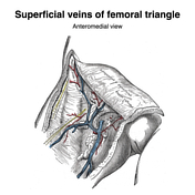
Superficial veins of the lower limb (Gray's illustration)
Published
08 Dec 2021
35% complete
Diagram
Case
Veins of the axilla (Gray's illustration)

Published
08 Dec 2021
35% complete
Diagram
Case
Vertebral venous plexuses (Gray's illustrations)

Published
08 Dec 2021
32% complete
Diagram
Case
Superficial veins of the elbow (Gray's illustration)

Published
08 Dec 2021
35% complete
Diagram
Case
Superficial veins of the hand (Gray's illustration)

Published
08 Dec 2021
35% complete
Diagram
Case
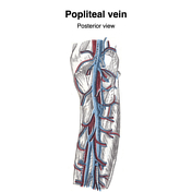
Popliteal vein (Gray's illustration)
Published
08 Dec 2021
32% complete
Diagram
Case
Anal triangle (diagrams)

Published
27 Oct 2021
35% complete
Diagram
Case
Urogenital triangle (diagrams)

Published
27 Oct 2021
35% complete
Diagram
Case
Female perineal muscles (Gray's illustration)

Published
26 Oct 2021
35% complete
Diagram
Case
Male perineal muscles (Gray's illustration)

Published
26 Oct 2021
35% complete
Diagram
Case
Male perineal fascia (Gray's illustration)

Published
26 Oct 2021
29% complete
Diagram
Case
Male pelvic and perineal fascia (Gray's illustrations)

Published
26 Oct 2021
29% complete
Diagram
Case
Levator ani (Gray's illustration)

Published
26 Oct 2021
32% complete
Diagram
Case
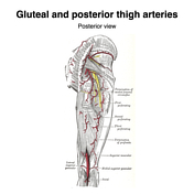
Gluteal arteries (Gray's illustration)
Published
26 Aug 2021
35% complete
Diagram
Case
Leg arteries (Gray's illustrations)

Published
26 Aug 2021
35% complete
Diagram
Case
Femoral artery (Gray's illustrations)

Published
26 Aug 2021
35% complete
Diagram
Case
Plantar arteries (Gray's illustrations)

Published
26 Aug 2021
32% complete
Diagram
Case
Genicular arteries (Gray's illustration)

Published
26 Aug 2021
32% complete
Diagram
Case
Obturator internus (Gray's illustration)

Published
25 Aug 2021
32% complete
Diagram
Case
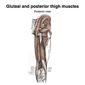
Gluteal and posterior thigh muscles (Gray's illustration)
Published
25 Aug 2021
35% complete
Diagram
Case
Medial thigh muscles (Gray's illustration)

Published
25 Aug 2021
32% complete
Diagram
Case
Saphenous hiatus (Gray's illustration)

Published
25 Aug 2021
35% complete
Diagram
Case
Anterior thigh muscles (Gray's illustration)

Published
25 Aug 2021
32% complete
Diagram
Case
Posterior shoulder muscles (Gray's illustration)

Published
25 Aug 2021
32% complete
Diagram
Case
Palmar aponeurosis (Gray's illustration)

Published
24 Aug 2021
35% complete
Diagram
Case
Thenar muscles (Gray's illustration)

Published
24 Aug 2021
35% complete
Diagram
Case
Intrinsic hand muscles (Gray's illustration)

Published
24 Aug 2021
35% complete
Diagram
Case
Hand interossei muscles (Gray's illustrations)

Published
24 Aug 2021
35% complete
Diagram
Case
Tendon sheaths of the wrist and hand (Gray's illustrations)

Published
24 Aug 2021
29% complete
Diagram
Case
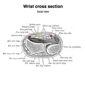
Wrist cross section diagrams (Gray's illustrations)
Published
18 Aug 2021
35% complete
Diagram
Case
Supinator (Gray's illustration)

Published
18 Aug 2021
32% complete
Diagram
Case
Posterior forearm muscles (Gray's illustrations)

Published
18 Aug 2021
32% complete
Diagram
Case
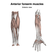
Anterior forearm muscles (Gray's illustrations)
Published
18 Aug 2021
32% complete
Diagram
Case
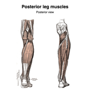
Posterior leg muscles (Gray's illustrations)
Published
15 Aug 2021
35% complete
Diagram
Case
Anterior and lateral leg muscles (Gray's illustration)

Published
15 Aug 2021
35% complete
Diagram
Case
Pectoral muscles (Gray's illustrations)

Published
15 Aug 2021
35% complete
Diagram
Case
Anterior abdominal wall (Gray's illustrations)

Published
15 Aug 2021
35% complete
Diagram
Case
Inguinal canal and rings (Gray's illustrations)

Published
03 Aug 2021
32% complete
Diagram
Case
Ankle tendons (Gray's illustrations)

Published
03 Aug 2021
35% complete
Diagram
Case
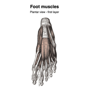
Plantar foot muscles (Gray's illustrations)
Published
03 Aug 2021
29% complete
Diagram
Case
Foot interossei muscles (Gray's illustrations)

Published
02 Aug 2021
35% complete
Diagram
Case
Rectus sheath (Gray's illustrations)

Published
02 Aug 2021
32% complete
Diagram
Case
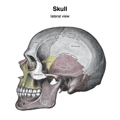
Skull (Gray's illustrations)
Published
18 May 2021
35% complete
Diagram
Case
Deep anterior chest wall muscles (Gray's illustration)

Published
12 May 2021
32% complete
Diagram
Case
Deep back muscles (Gray's illustration)

Published
12 May 2021
35% complete
Diagram
Case
Deep neck muscles (Gray's illustration)

Published
12 May 2021
35% complete
Diagram
Case
Hyoid bone (Gray's illustration)

Published
05 May 2021
32% complete
Diagram
Case
Levine and Edwards classification of hangman fractures (diagrams)

Published
06 Apr 2021
35% complete
Diagram
Case
Gehweiler classification of atlas fractures (diagrams)

Published
11 Mar 2021
35% complete
Diagram
Case
Roy-Camille classification of C2 odontoid fractures (diagrams)

Published
03 Mar 2021
35% complete
Diagram
Case
Anderson and D'Alonzo classification of C2 odontoid fractures (diagrams)

Published
25 Feb 2021
35% complete
Diagram
Case
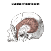
Muscles of mastication (Gray's illustration)
Published
24 Feb 2021
35% complete
Diagram
Case
Anderson and Montesano classification of occipital condyle fractures (diagrams)

Published
23 Feb 2021
35% complete
Diagram
Case
Traynelis classification of atlanto-occipital dissociation (diagrams)

Published
23 Feb 2021
35% complete
Diagram
Case
Thoracolumbar injury classification and severity score (table)

Published
16 Feb 2021
10% complete
Diagram
Case
Subaxial cervical spine injury classification (table)

Published
16 Feb 2021
10% complete
Diagram
Case
Lymphatics of the thorax and abdomen (Gray's illustration)

Published
25 Jan 2021
29% complete
Diagram
Case
Lymphatics of the lower limb (Gray's illustration)

Published
29 Dec 2020
35% complete
Diagram
Case
Lymphatics of the popliteal fossa (Gray's illustration)

Published
29 Dec 2020
35% complete
Diagram
Case
Ankle and foot interosseous ligaments (Gray's illustrations)

Published
20 Dec 2020
32% complete
Diagram
Case
Lymphatics of the upper limb (Gray's illustration)

Published
20 Dec 2020
35% complete
Diagram
Case
Plantar ligaments of the foot (Gray's illustration)

Published
17 Dec 2020
35% complete
Diagram
Case
Lateral talocrural ligaments (Gray's illustration)

Published
17 Dec 2020
35% complete
Diagram
Case
Ankle and foot ligaments (Gray's illustrations)

Published
17 Dec 2020
35% complete
Diagram
Case
Subtalar ligaments (Gray's illustration)

Published
17 Dec 2020
35% complete
Diagram
Case
Patella (Gray's illustration)

Published
14 Dec 2020
32% complete
Diagram
Case
Knee joint capsule (Gray's illustrations)

Published
14 Dec 2020
35% complete
Diagram
Case
Hoffa's fat pad (Gray's illustration)

Published
14 Dec 2020
32% complete
Diagram
Case
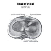
Knee menisci (Gray's illustrations)
Published
10 Dec 2020
35% complete
Diagram
Case
Internal knee ligaments (Gray's illustrations)

Published
10 Dec 2020
35% complete
Diagram
Case
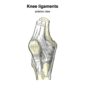
Knee ligaments (Gray's illustrations)
Published
10 Dec 2020
35% complete
Diagram
Case
Hip joint capsule (Gray's illustration)
Published
02 Dec 2020
35% complete
Diagram
Case
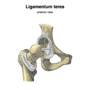
Ligamentum teres of the hip (Gray's illustrations)
Published
02 Dec 2020
35% complete
Diagram
Case
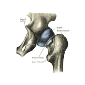
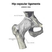
Hip capsular ligaments (Gray's illustrations)
Published
30 Nov 2020
35% complete
Diagram
Case
Ligaments of the fingers (Gray's illustrations)

Published
30 Nov 2020
35% complete
Diagram
Case
Triangular fibrocartilage complex (Gray's illustration)
Published
30 Nov 2020
32% complete
Diagram
Case
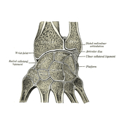
Wrist ligaments (Gray's illustrations)

Published
25 Nov 2020
32% complete
Diagram
Case
Proximal radio-ulnar joint ligaments (Gray's illustration)

Published
24 Nov 2020
35% complete
Diagram
ADVERTISEMENT: Supporters see fewer/no ads


 Unable to process the form. Check for errors and try again.