
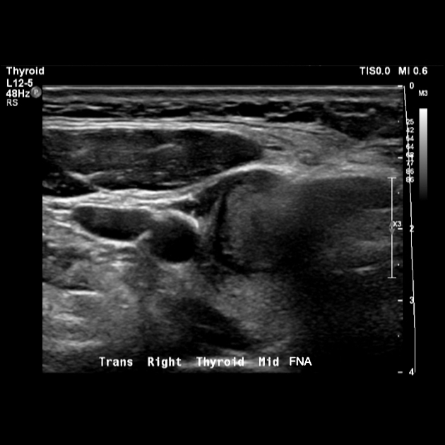
Ultrasound is the first-line imaging modality for assessment of thyroid nodules found on clinical examination or incidentally on another imaging modality. This article is an overview of ultrasonographic features of thyroid nodules, which are used to determine the need for biopsy with fine needle aspiration (FNA).
Following concerns regarding variation in practice and overdiagnosis of clinically insignificant malignancy several professional societies have published formal assessment criteria to determine the need for FNA which are covered in separate articles.
On this page:
Radiographic features
Ultrasound
Calcification
Although calcification can be seen in both benign and malignant processes, it is the ultrasound feature most closely associated with malignancy 1.
-
microcalcifications
-
punctate echogenic foci without posterior shadowing
often might not actually represent calcifications 6
most specific finding associated with malignancy (~95%) 2
associated with papillary thyroid carcinoma
colloid (in benign colloid nodules) shows comet tail artifact (not ring-down); if an echogenic focus is not definitely colloid, biopsy is warranted
-
-
coarse calcifications
can be seen in both benign and malignant nodules
associated with both papillary thyroid carcinoma and medullary thyroid carcinoma
-
peripheral rim calcification
can be seen in both benign and malignant nodules
Echogenicity
-
hypoechoic solid nodule
most papillary thyroid carcinomas
nearly all medullary thyroid carcinomas 3
benign nodules can be hypoechoic
if no other malignant features (e.g. calcifications) then hypoechoic nodules are typically biopsied after reaching size criteria
isoechoic solid nodule: 25% (follicular and medullary)
hyperechoic solid nodule: 5% chance of being malignant
large cystic component favors a benign entity although a significant proportion of papillary carcinomas will have a cystic component
while a halo around a well-marginated hypoechoic or isoechoic nodule is typical of a follicular adenoma 3, it is absent in >50% of benign nodules 2; what is more, up to 24% of papillary thyroid carcinomas may have a halo, be it complete or incomplete 2
Color Doppler
intranodular flow usually malignant
lymph nodes with increased color Doppler flow are suspicious
Other
invasion of local structures favors anaplastic thyroid carcinoma and thyroid lymphoma
shadowing around the edges of a nodule (edge refraction shadow) are associated with papillary thyroid carcinoma 3
a nodule taller than it is wide is suspicious for malignancy 4
irregular margins are suspicious for malignancy 4
Lymph nodes
enlarged regional lymph nodes are suspicious for thyroid malignancy, especially papillary thyroid carcinoma
microcalcifications in regional lymph nodes are highly suspicious
lymph nodes with cystic change are highly suspicious
loss of normal fatty hilum, irregular node appearance
increased color Doppler flow is suspicious
-
no threshold criteria for lymph node biopsy
biopsy if suspicious features
consider biopsy if >8 mm
Sonographic features favoring a benign nodule
large cystic component
hyperechoic solid
comet tail artifact
spongiform appearance / sponge-like appearance 7,8
Sonographic features favoring a malignant nodule
hypoechoic solid
presence of microcalcifications: almost always warrants biopsy
local invasion of surrounding structures
taller than it is wide
large size: the cutoff is often taken as 10 mm to warrant biopsy
suspicious neck lymph nodes suggesting metastatic disease
intranodular blood flow
Differential diagnosis
The differential for a suspicious nodule includes benign nodules such as adenomatoid nodules, follicular adenoma, and Hashimoto thyroiditis. Parathyroid adenomas are also confounding nodules.
No sonographic features are 100% sensitive or specific (although lymphadenopathy with microcalcifications is 100% specific). A suspicious nodule (and lymph node, if applicable) should undergo biopsy with fine needle aspiration. Multiple criteria exist from societies in different subspecialties and biopsy thresholds vary among institutions. See below for individual systems.


 Unable to process the form. Check for errors and try again.