
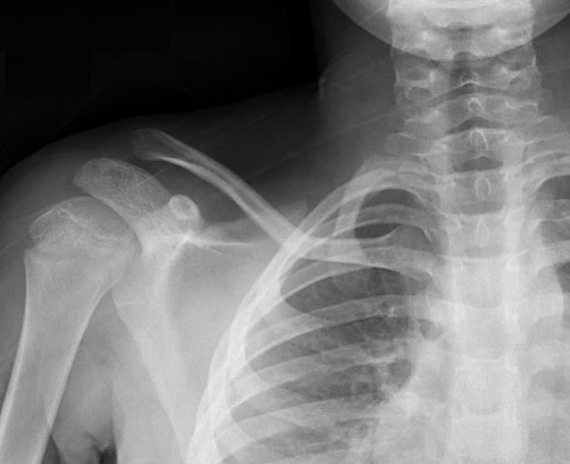
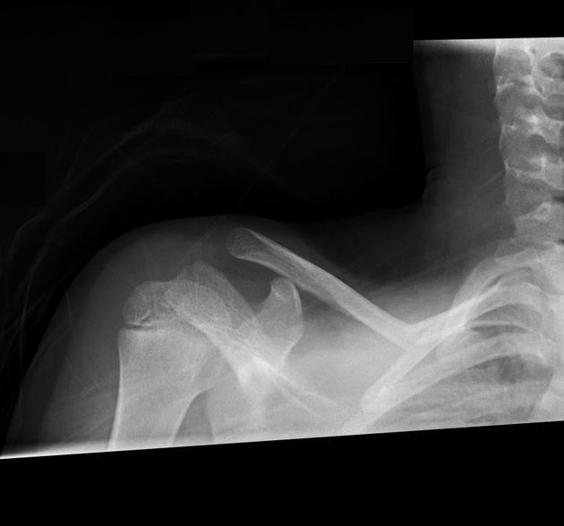
The clavicle series for pediatrics is a two-view series containing an anteroposterior and a cranially angled axial radiograph. Depending on the patient's level of distress and severity of the injury, adapting the radiographic technique to suit a child sitting in bed or lying supine may be necessary.
On this page:
Indications
In pediatrics, clavicle radiographs are mainly indicated for:
trauma with a suspected fracture
acromioclavicular or sternoclavicular dislocation
Projections
Standard projections
Patient preparation
Patients should remove any clothing, arm sling or jewelry from the affected clavicle.
Gonadal shielding
The use of gonadal and fetal shielding has been deemed as non-beneficial to patients' health per current evidence and may or may not be useful for pediatric extremity imaging 1-3. The use of gonadal shielding can increase the examination time and may cause the child more distress.
Tips for pediatric clavicle radiography
The major difficulty in pediatric radiography of the clavicle relates to:
To overcome this, a variety of techniques can be used:
distract the patient with toys, games and/or conversation
using the swaddling technique; wrap the child in a blanket to promote comfort and perform the images supine
setting up the room before moving the patient into the required position can reduce patient distress
Immobilization techniques
Children will find it difficult to keep their arm still; particularly if the limb is injured. One option is to have a carer or radiographer stand on the child's non-affected side, placing one hand on their mid-chest and another holding the proximal forearm.


 Unable to process the form. Check for errors and try again.