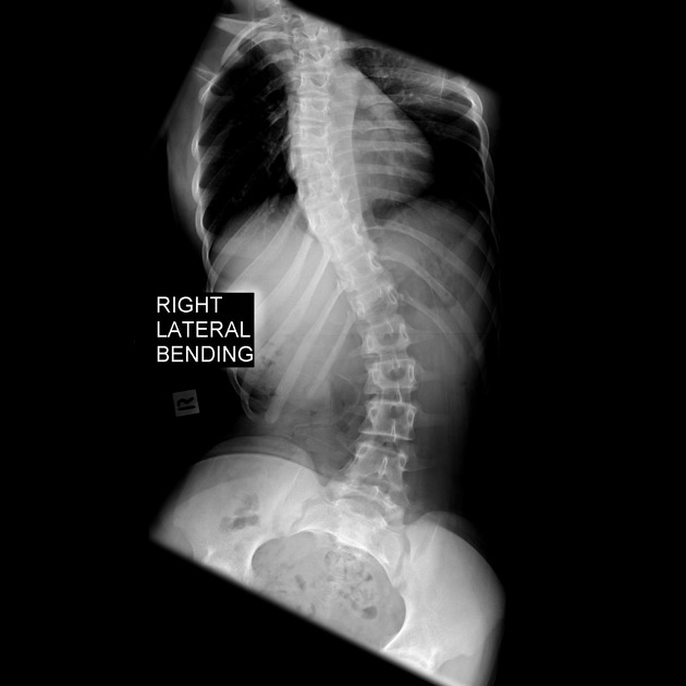
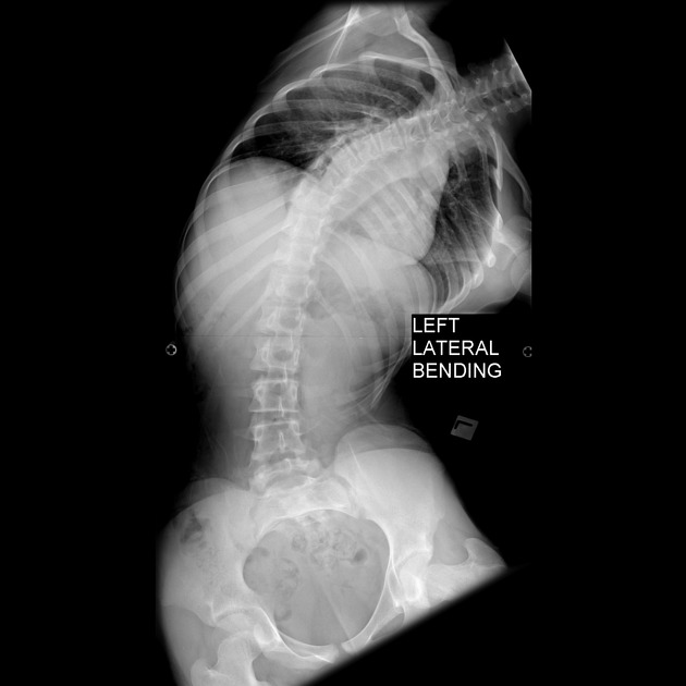
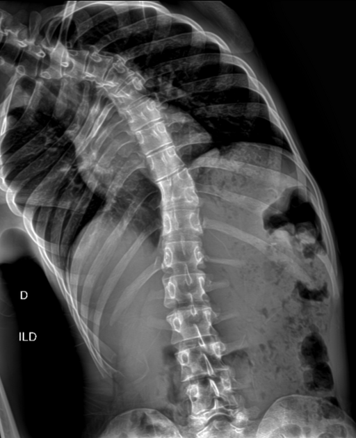
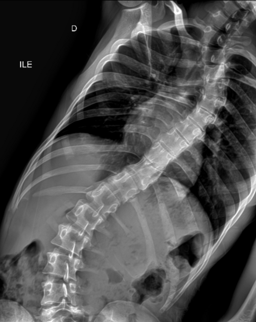
Scoliosis lateral bending views are additional scoliosis projections accompanying the standard PA/AP and lateral views.
On this page:
Indications
The aim of this view is to assess patients' lateral range of spinal motion 1 in the vertebral column as part of a scoliosis series.
Patient position
patient erect or supine
patient bending their upper body laterally (right and left) from the hips as much as possible
ensure patient aligned centrally to the IR
ensure rotation of hips and shoulders is reduced as much as possible (some rotation inherent to scoliosis may be inevitable)
ensure at least 3-5 cm of iliac crests are present on the radiograph
Technical factors
anteroposterior or posteroanterior projection
suspended expiration
-
centering point
dependent on the area of interest, patient height and detector size
-
collimation
dependent on orthopedic preference and imaging modality
superiorly to include all vertebrae of interest (may be at C7)
inferiorly to include the sacral region (may be at S1 or level of femoral heads)
lateral collimation sufficient to include all of spinal curvature
-
orientation
portrait
-
detector size
most likely a stiched series
-
exposure
80-95 kVp (digital)
40-60 mAs
-
SID
100-150 cm
-
grid
yes
Image technical evaluation
area of scoliosis should be visible with evidence of iliac crests inferiorly
-
no patient rotation
-
rotated vertebrae may be distinguished from scoliotic vertebrae in that:
rotated vertebral bodies will have their long axes straight and
scoliotic vertebral bodies will have a lateral deviation
-
bony margins and trabecular patterns should both be clearly visible in thoracic and lumbar vertebrae
Practical points
-
PA projections should be considered over the AP projection for the reduced dose to radiosensitive organs situated anteriorly
although the PA projection may be difficult to achieve if the patient is in a prone position
a compensatory wedge filter may be appropriate to achieve isodensity throughout the image 1
-
certain x-ray systems have automatic tracking functions to scan the entire vertebral column in two to three separate images before a post-processing stitch is applied
as it may be uncomfortable for patients to remain in the lateral bending position for the duration of such procedures, inform them beforehand of the duration, and ensure you instruct them to stand absolutely still as any slight movement may affect the stitching process afterwards
inform the patient once the image is acquired so that they can stand upright again


 Unable to process the form. Check for errors and try again.