
Crossed fused renal ectopia refers to an anomaly where the kidneys are fused and located on the same side of the midline.
On this page:
Epidemiology
The estimated incidence is around 1 out of 1000 births 1. There is a recognized male predilection with a 2:1 male to female ratio. More than 90% of crossed renal ectopia results in fusion.
Pathology
-
cause:
results from abnormal renal ascent during embryogenesis, with fusion of the kidneys occurring in the pelvis
occurs during the 4th-8th week of fetal life (normal renal ascent to l2 level is complete by the end of the second month)
-
theories of pathogenesis:
-
umbilical artery hypothesis
an abnormally positioned umbilical artery impedes normal cephalic migration of the kidneys
-
ureteric bud hypothesis
the ureteric bud crosses to the opposite side, inducing nephron formation in the contralateral metanephric blastema
-
-
outcome:
results in a single renal mass with two collecting systems located on one side of the abdomen
-
effects on fascial development:
normal kidney ascent is necessary for the formation of extraperitoneal perirenal fascial planes
in ectopia (or renal agenesis), there is a failure of fascial development in the flank on the side without renal tissue
-
consequences include:
bowel malposition: loops of bowel may occupy the extraperitoneal fat in the empty renal fossa
relaxation of mesenteric supports: increased mobility of bowel loops in the region of the empty renal fossa
Subtypes
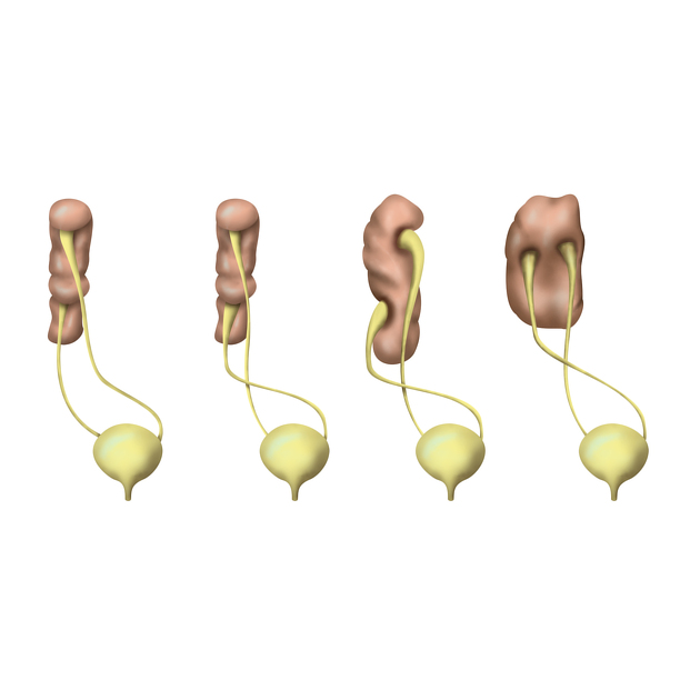
They are subclassified into six subtypes in decreasing order of frequency 6
type a: inferior crossed fusion
type b: sigmoid kidney
type c: lump kidney
type d: disc kidney
type e: L-shaped kidney
type f: superiorly crossed fused
Location
Left-to-right ectopy is thought to be three times more common.
Radiographic features
Fluoroscopy
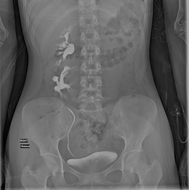
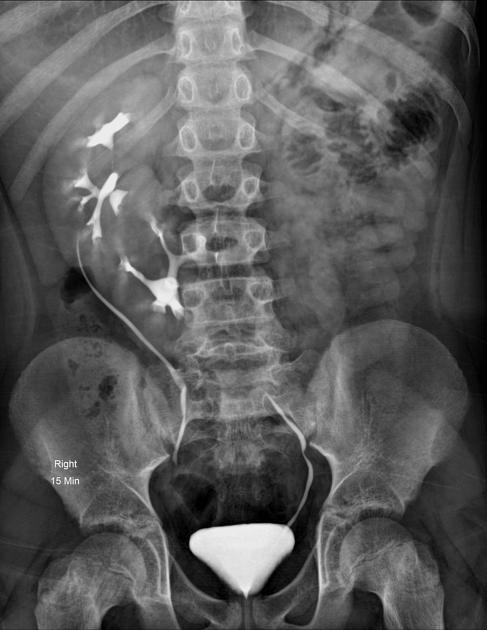
Urography (IVU)
The anomaly is readily detected on conventional urography. In 90% of crossed ectopy, there is at least partial fusion of the kidneys (the remainder demonstrate two discrete kidneys on the same side, crossed-unfused ectopy).
An anterograde or retrograde ureterogram most often demonstrates normal bladder trigone without ureteral ectopy.
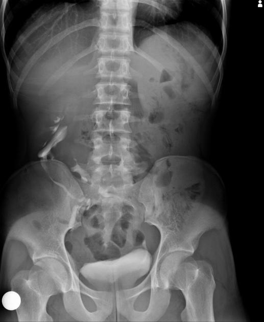
Barium studies of the bowel
Barium contrast studies of the bowel should be interpreted in light of bowel laxity in the region of the empty renal fossa (discussed above). In particular, a distinction must be made from internal hernia.
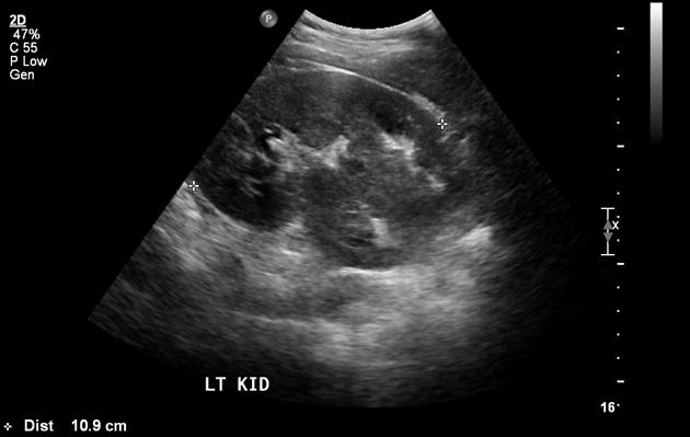
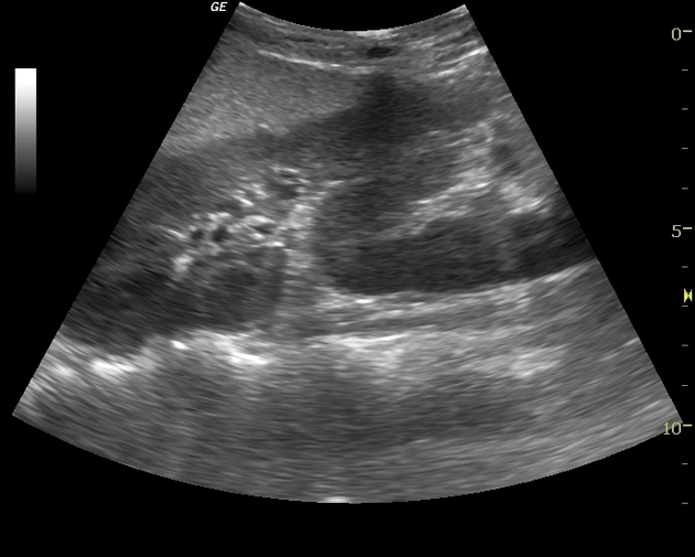
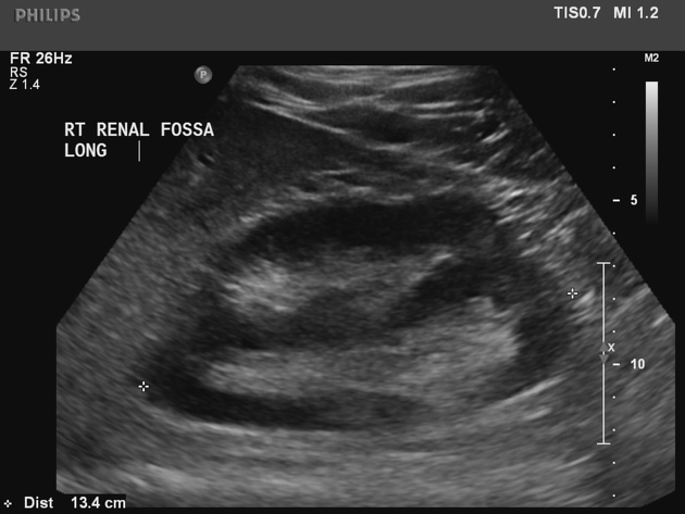
Ultrasound
On ultrasound, there may be a characteristic anterior or posterior "notch" between the two fused kidneys.
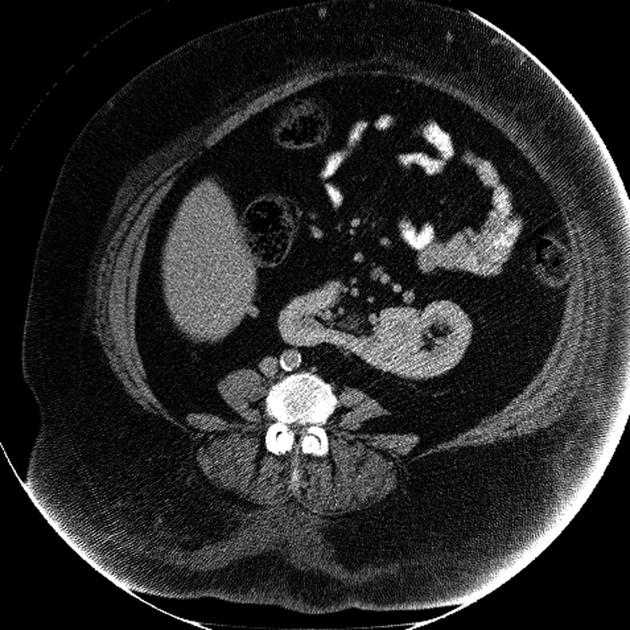
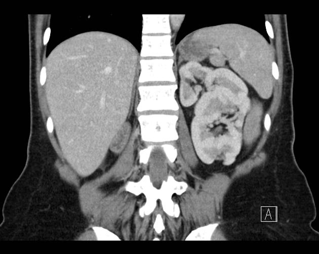
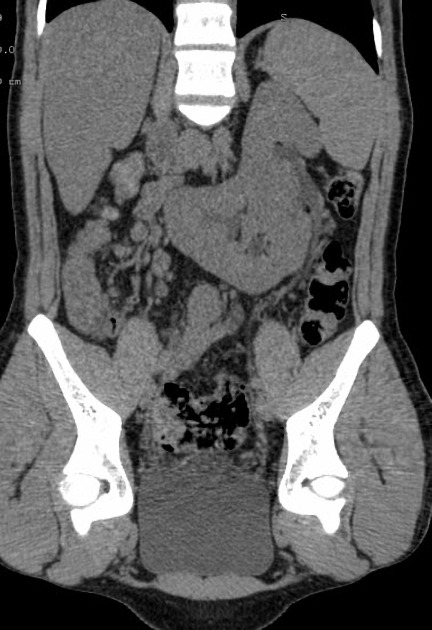
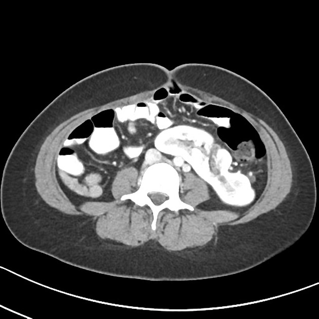
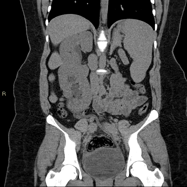
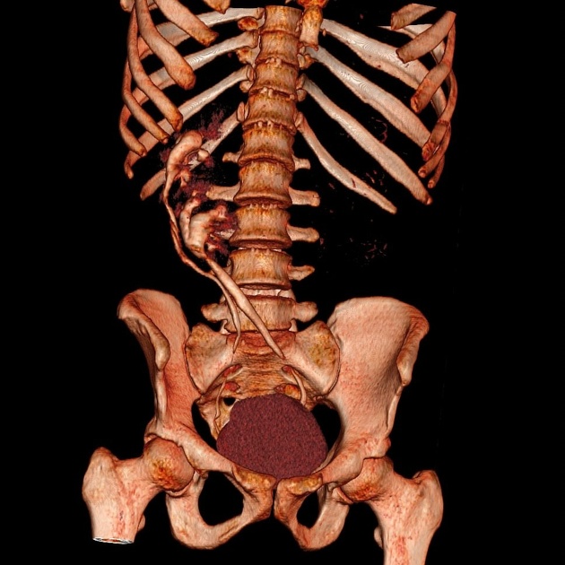
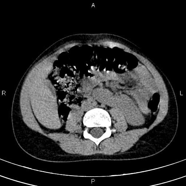
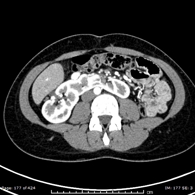
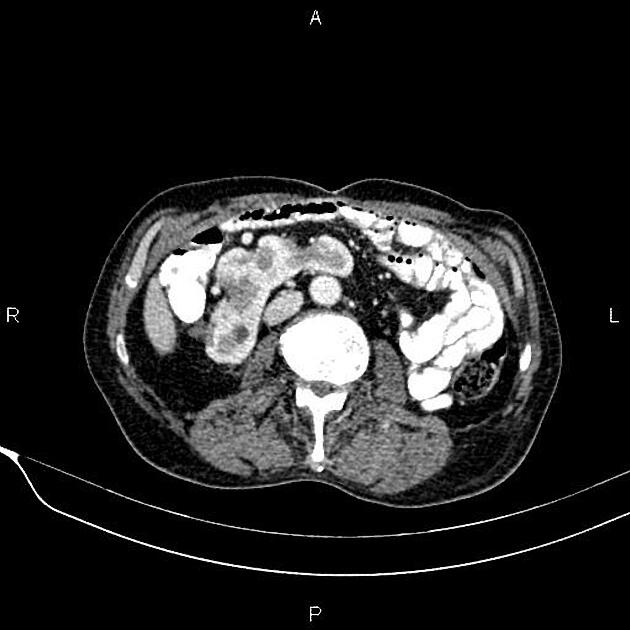
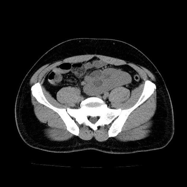
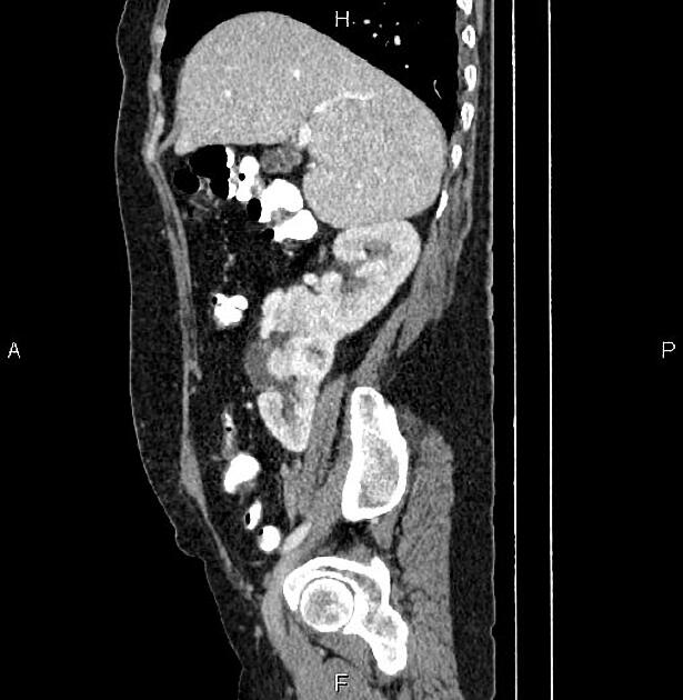
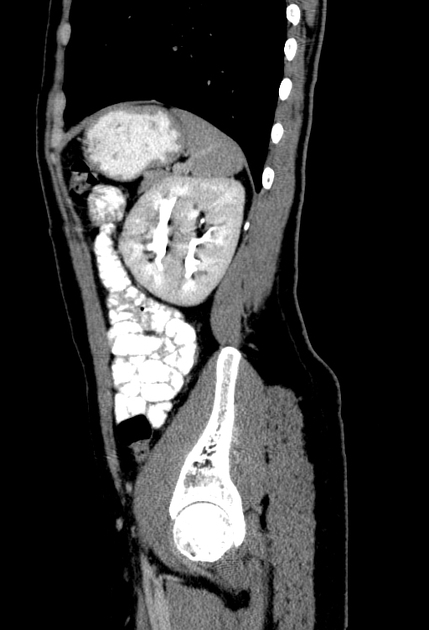
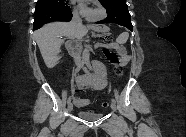
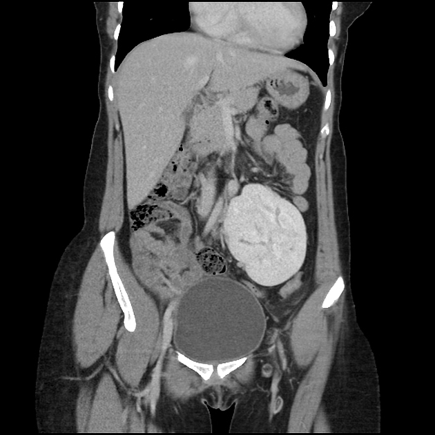
CT
The parenchymal band joining the two kidneys can be better visualized on CT scan. Also, anatomical relationship with adjacent structures and positions of the ureter can be better assessed.
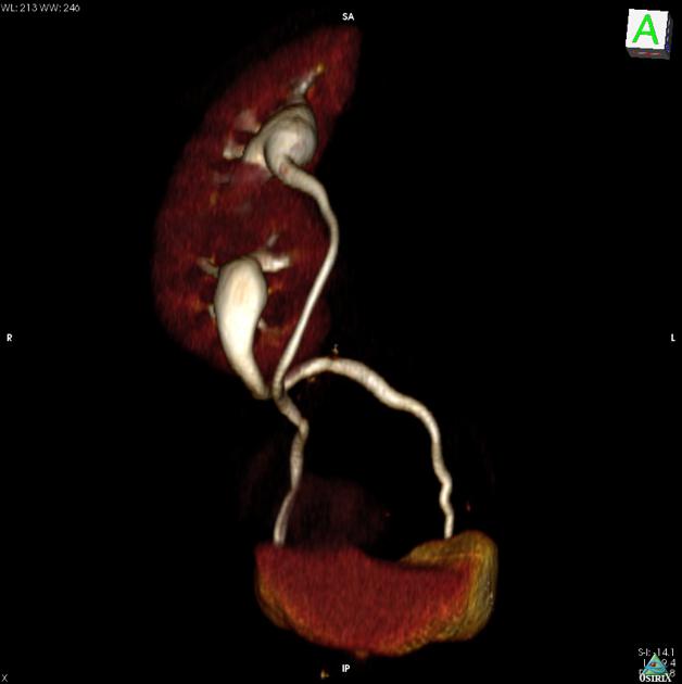
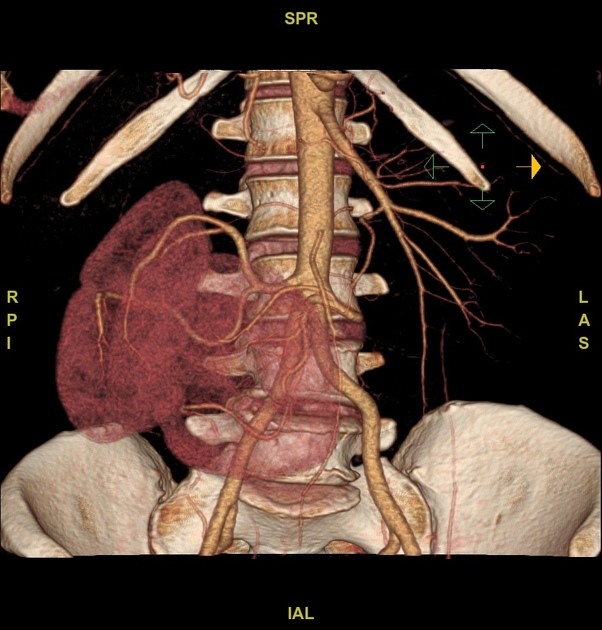
Treatment and prognosis
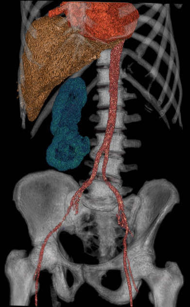
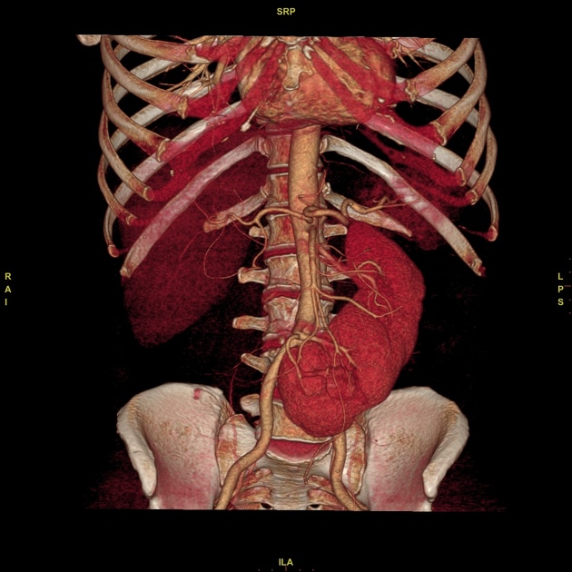
Crossed fused ectopia usually does not require any primary treatment. However, understanding is essential before planning any surgical intervention in the renal region. The blood supply to the crossed fused kidney is usually anomalous, and angiography is recommended before surgical intervention.
Complications
In a crossed fused renal ectopic kidney, complications such as nephrolithiasis, infection, and hydronephrosis approaches ~50%.
History and etymology
In 1654, Dominicus Panarolus was the first who described cross-fused renal ectopia.


 Unable to process the form. Check for errors and try again.